
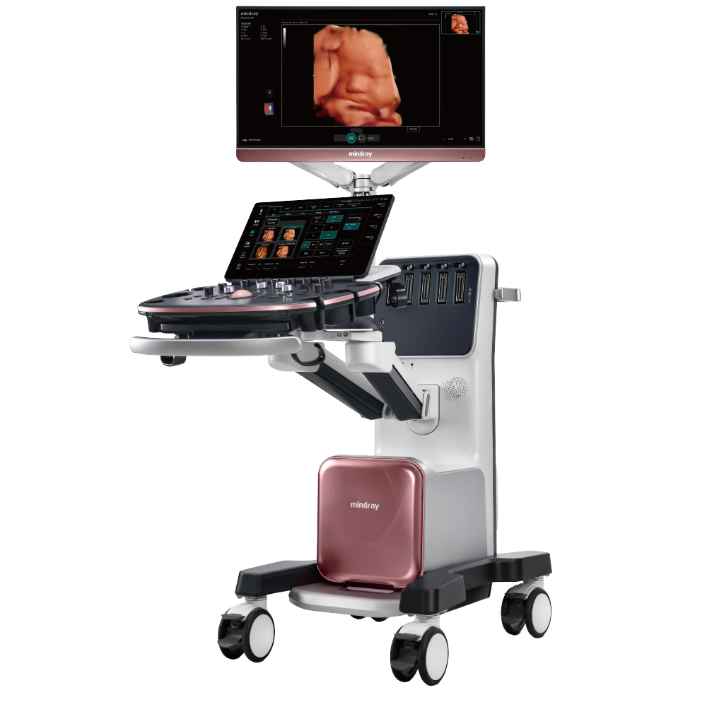
3x integrated computing power
High-throughput parallel processing ability
(powerful architecture (CPU, GPU))
Women's health ultrasound faces critical challenges in precision,
early detection, and standardized care. Introducing the
 , powered by the next-generation iZST+ platform evolved
from ZST+. With triple the computing power for high speed
parallel processing, approx. doubled acoustic-electric data quality
and N types of innovative algorithms that unlock new capabilities,
it enables clearer visualization of complex fetal structures,
earlier detection of subtle lesions, and more efficient high-quality
exams driven by full-stack intelligence - empowering clinicians to
work with greater confidence and turn infinite possibilities into
clinical reality.
, powered by the next-generation iZST+ platform evolved
from ZST+. With triple the computing power for high speed
parallel processing, approx. doubled acoustic-electric data quality
and N types of innovative algorithms that unlock new capabilities,
it enables clearer visualization of complex fetal structures,
earlier detection of subtle lesions, and more efficient high-quality
exams driven by full-stack intelligence - empowering clinicians to
work with greater confidence and turn infinite possibilities into
clinical reality.

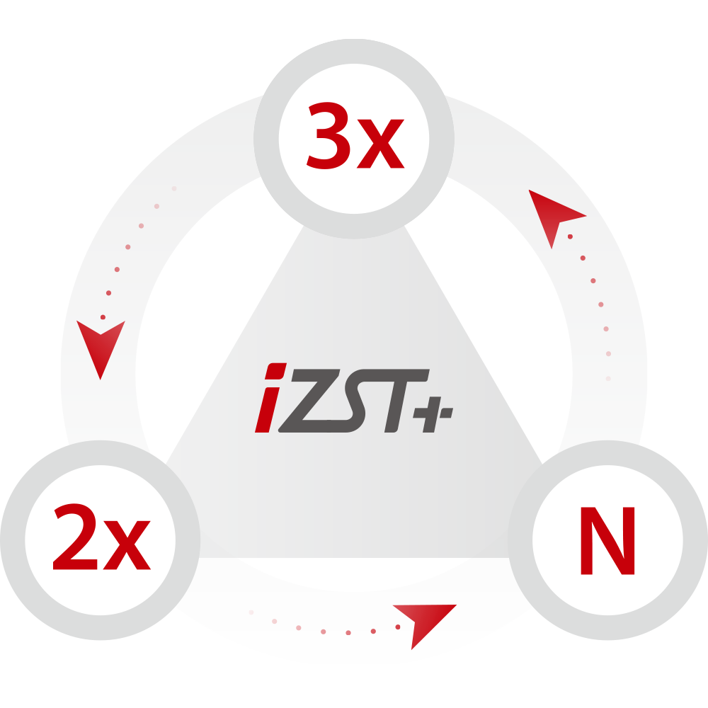

High-throughput parallel processing ability
(powerful architecture (CPU, GPU))

higher quality of acoustoelectric data for imaging
(premium-migrated technologies)

unlocking more possibilities and capabilities
(more imaging advancements)
From acoustic data acquisition at the transducer to pre and post imaging processing





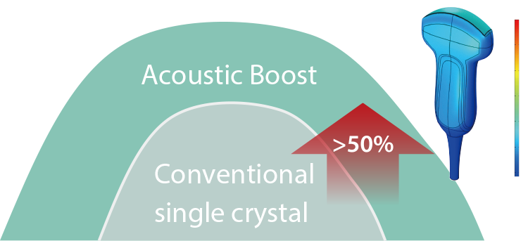
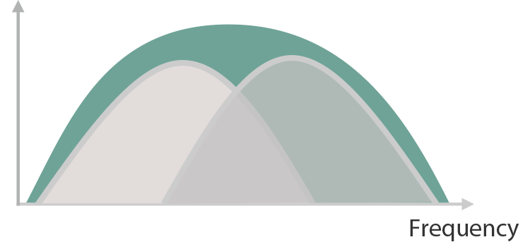
Premium-migrated technologies for acoustic data, doubling the acoustic signal quality of enriched datasets right from the start to achieve superb definition.
Enhanced signal strength, improved by over 50%
Wider bandwidth, increased by more
than 30%
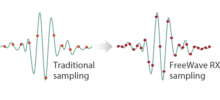
The sampling rate of the system is increased by 100%
Utilizes adaptive image analysis for acoustic shadow detection and suppression, effectively compensating for signal loss to restore obscured tissue details particularly behind strong echogenic structures (e.g., fetal skull or spine)

DeShadow on and off
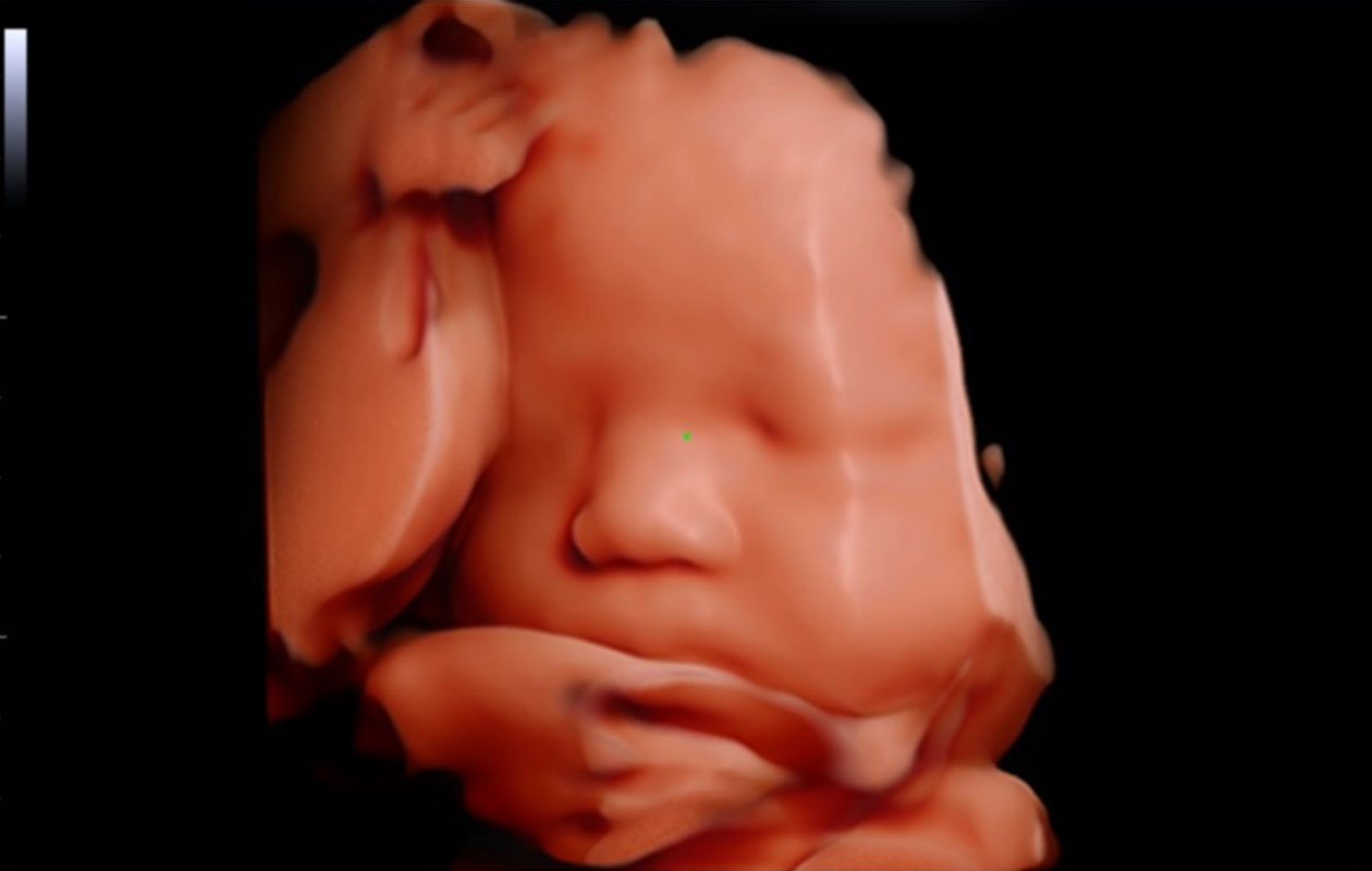
iLive with VR iTouch
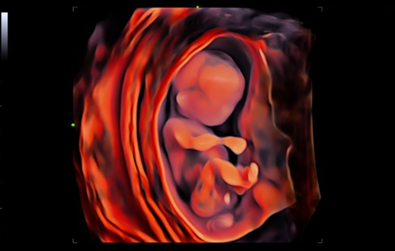
iLive with Hyaline (profile)
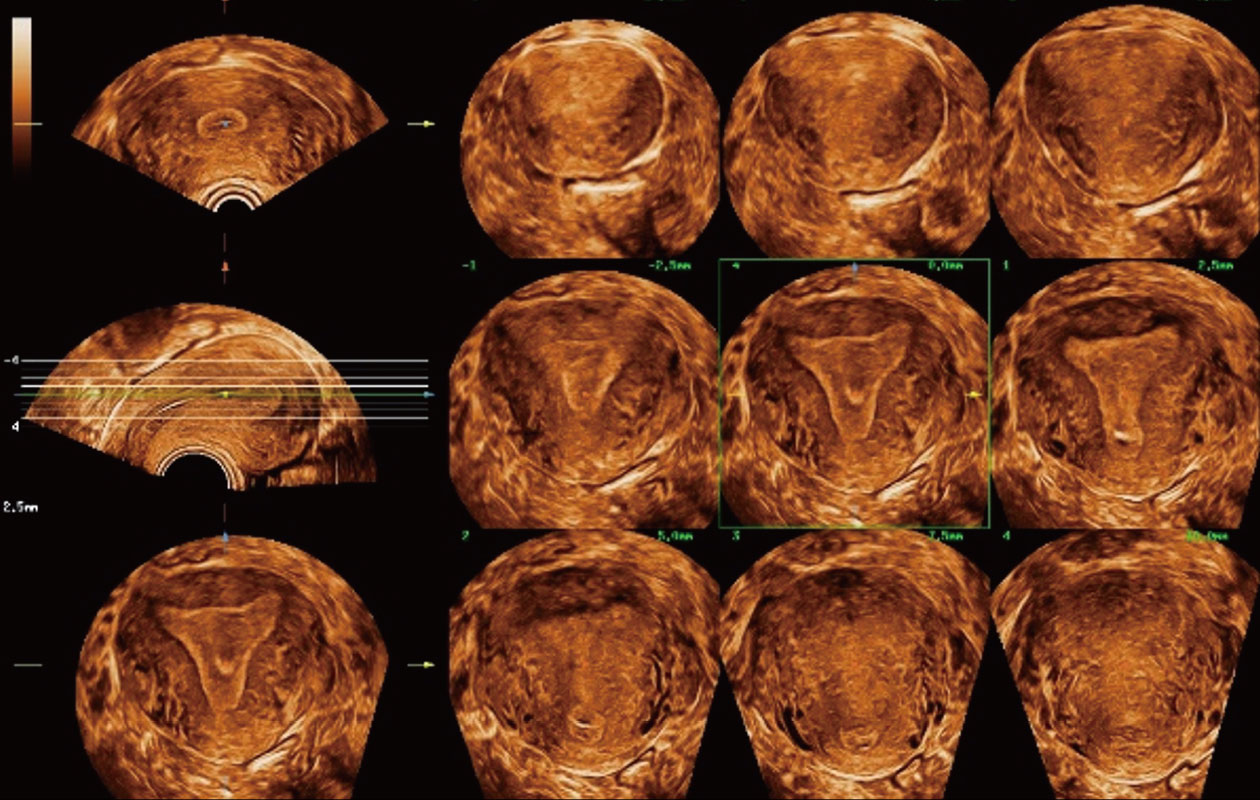
Endometrial structure details in morphology
Specialized clinical solutions with innovative insights for women's healthcare through entire life cycle










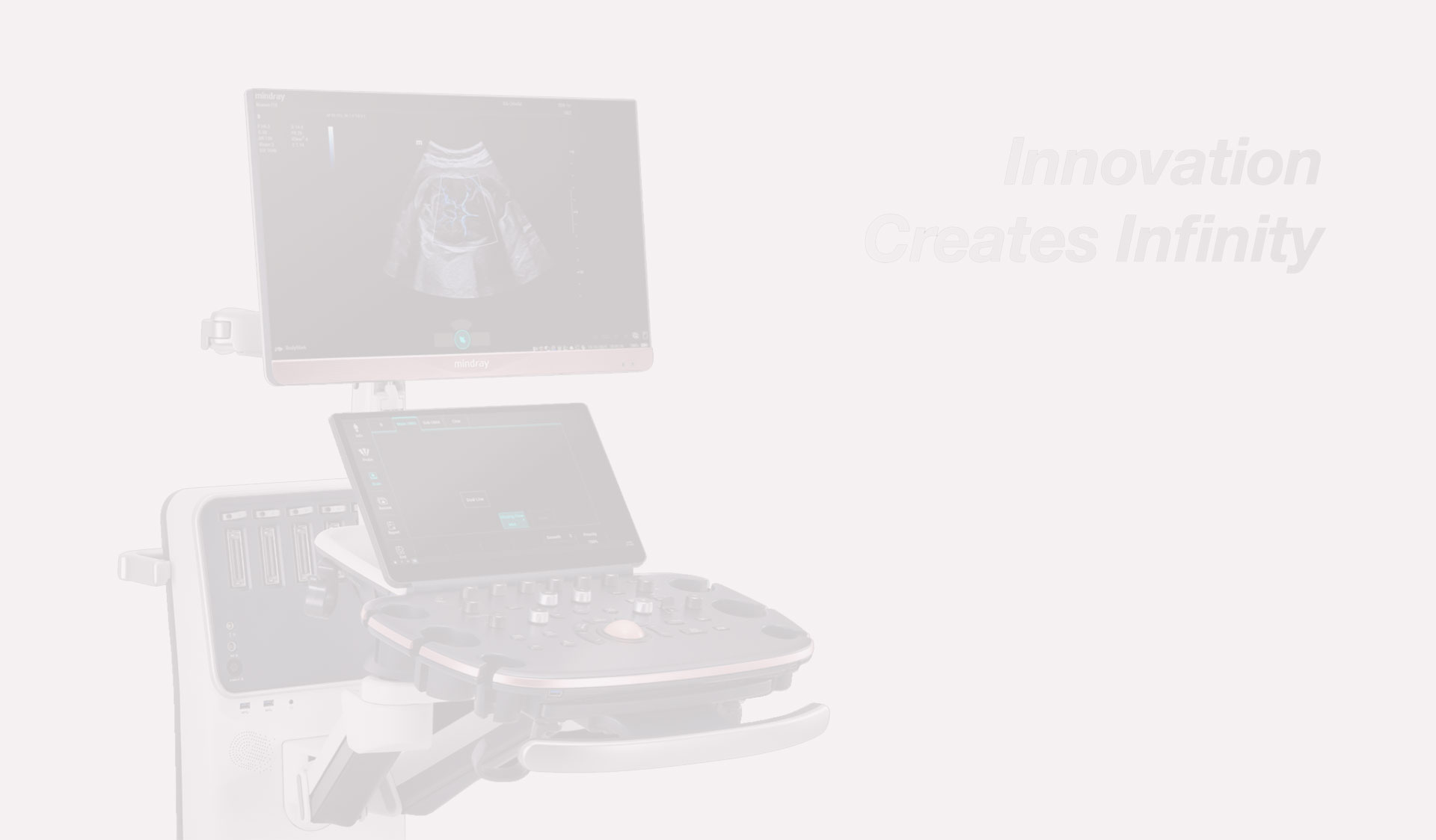
It enables full-stack intelligence based on obstetric scan protocols, encompassing anatomical structure recognition, standard plane acquisition, intelligent measurement, annotation, and quality control. For a complex prenatal screening exam, it can save an average of 10 minutes, reduce over 300 button clicks, and significantly enhance examination accuracy and consistency.
Smart OB Vue*
Standard plane recognition, storage, and quality control
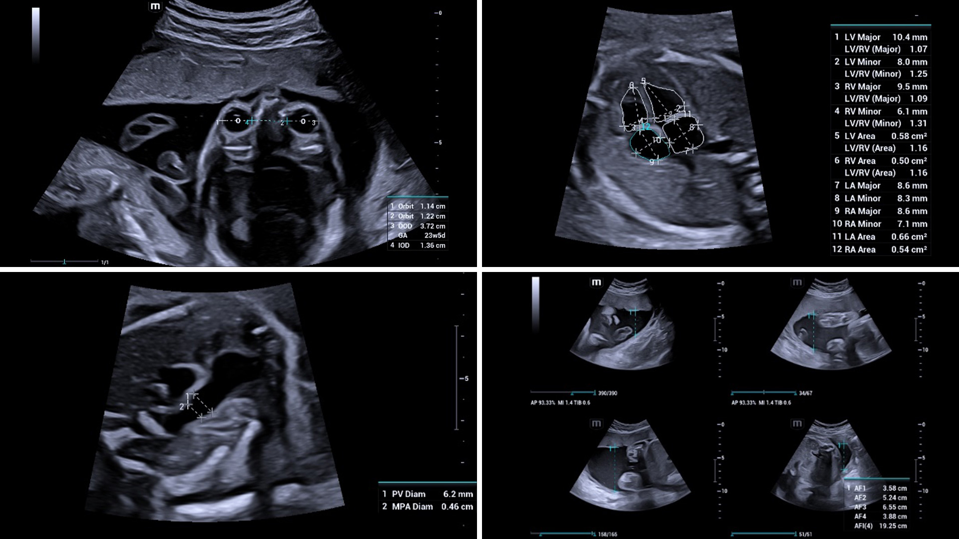
Smart OB Calc
Automatically identify and measure required measurements
* The availability of this function is subject to country-specific policies and regulations.
High-clarity images with subtle details and Smart Scene intelligence help to easily acquire the most significant planes and obtain frequently used measurements of fetal brain for smart diagnosis, enabling improved throughput and reduced user dependency.
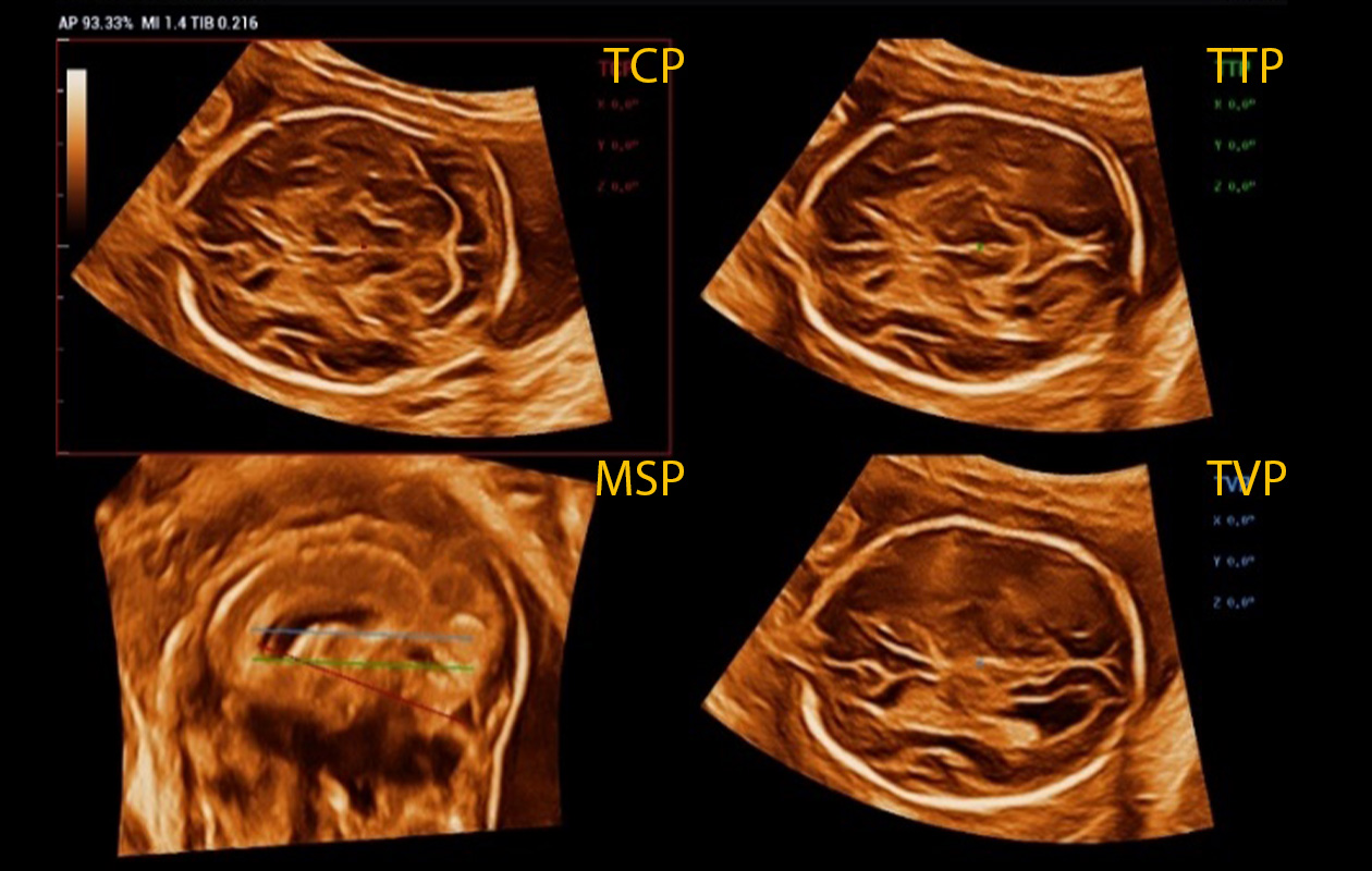
Smart Planes CNS
Full automatic planes acquisition and measurements
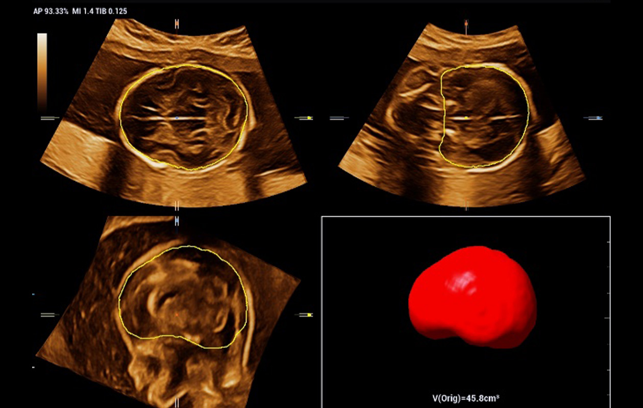
Smart ICV
Fully automatic intra-cranial volume calculation
Smart Planes Spine* reduces the difficulty in scanning and intelligently splits vertebral bodies and arches for clear observation and reduced misdiagnosis of fetal spina bifida. The guide line also enables much easier and more confident positioning of conus medullaris.
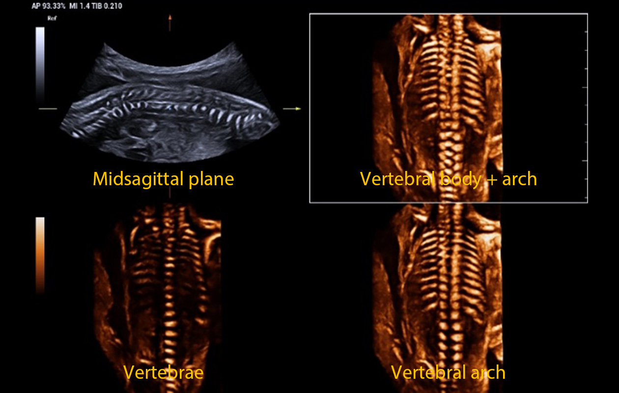
Smart Planes Spine*
Automatically separate the imaging of vertebral arch and body
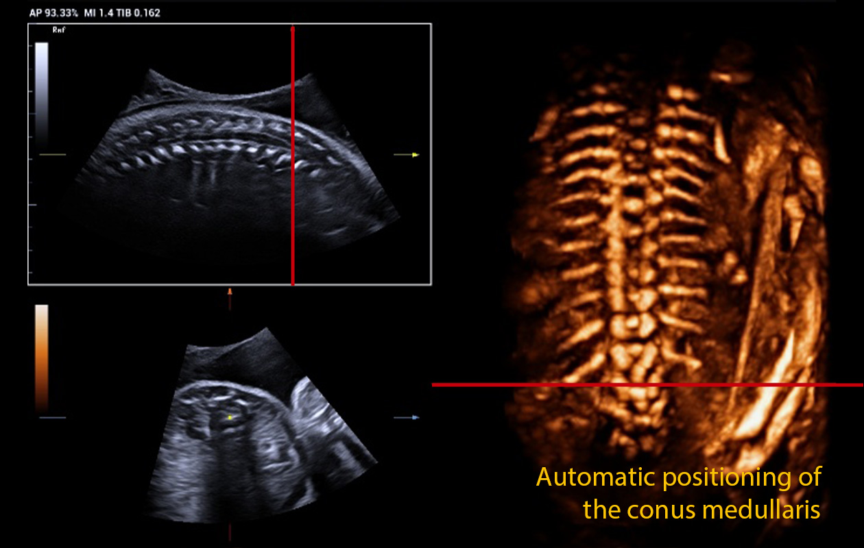
Smart Planes Spine*
Fully automatic positioning of the conus medullaris with the reference line
* The availability of this function is subject to country-specific policies and regulations.

Smart scene-endometrium realizes intelligent endometrial thickness measurement, volume rendering, auto coronal plane acquisition, and even 2D or 3D vascularity study for endometrial receptivity analysis.
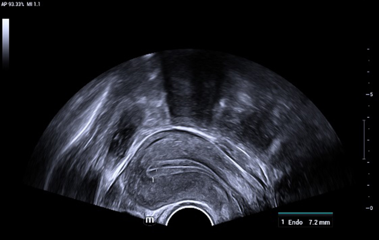
Auto measure of endometrial thickness
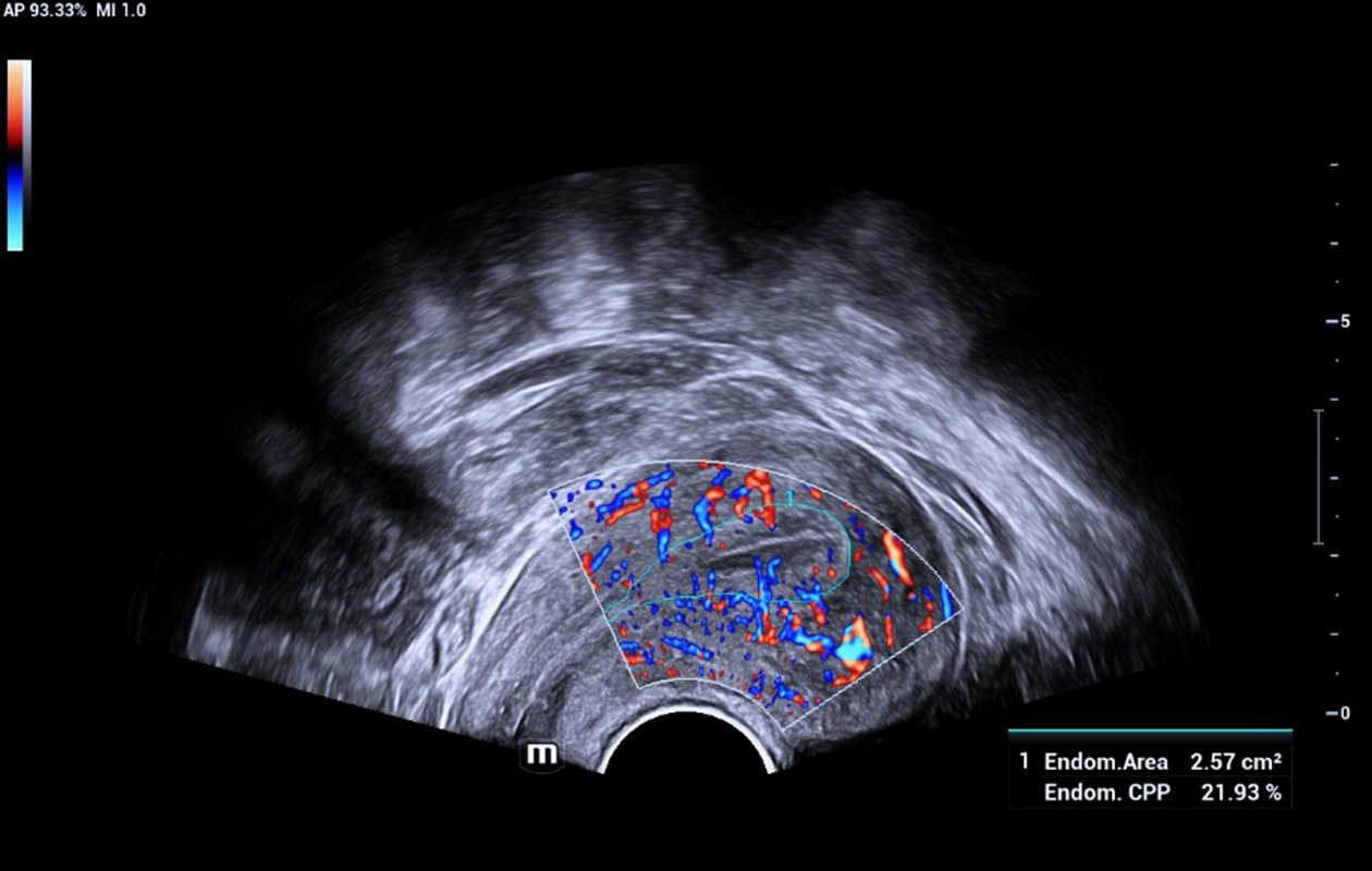
Auto tracing and flow analysis of endometrium
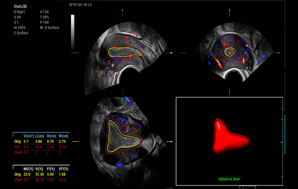
Auto endometrial volume rendering and quantification
The brand new Smart FLC provides precise volume rendering, count and quantitative analysis of ovary and even antral follicles for easy, effective and confident ovarian reserve assessment.
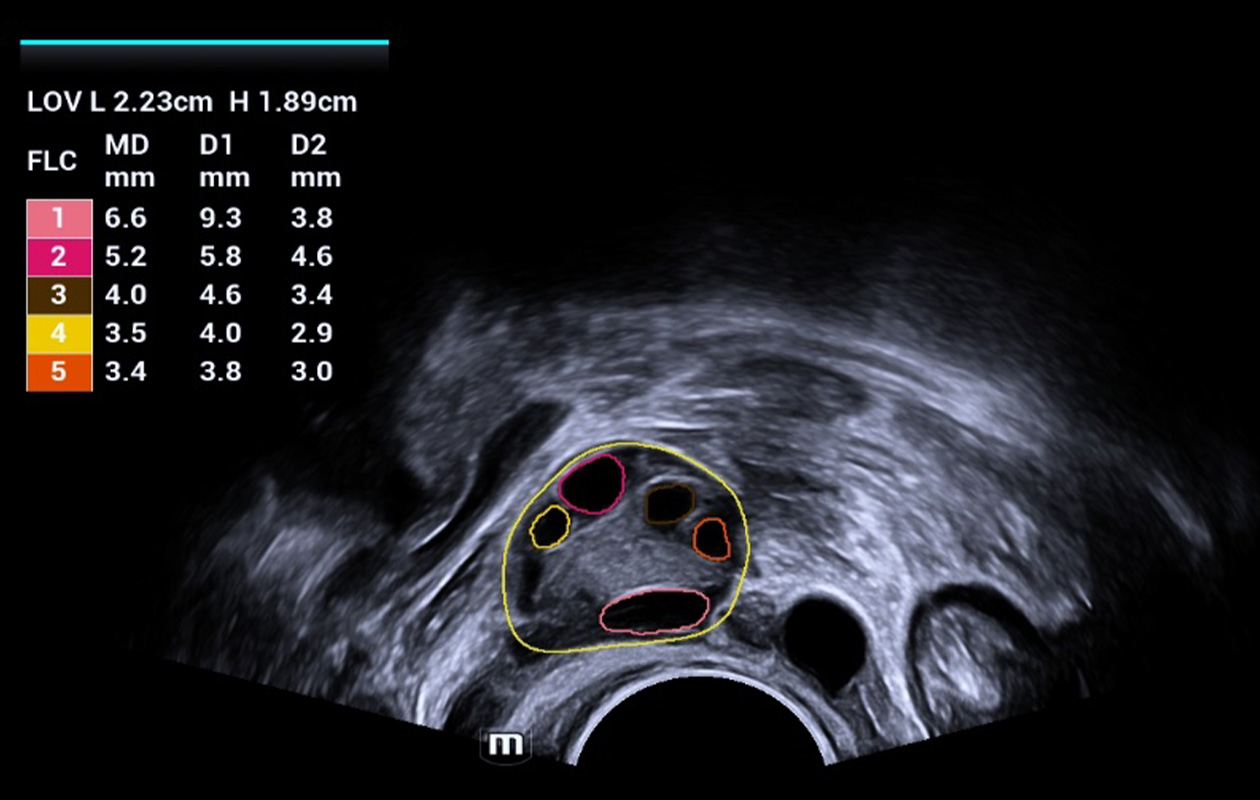
Auto measure of endometrial thickness
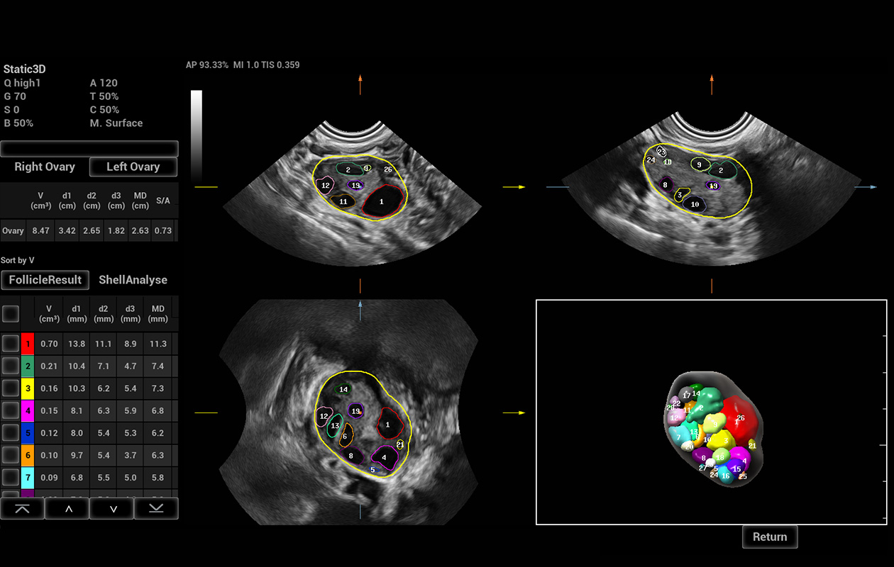
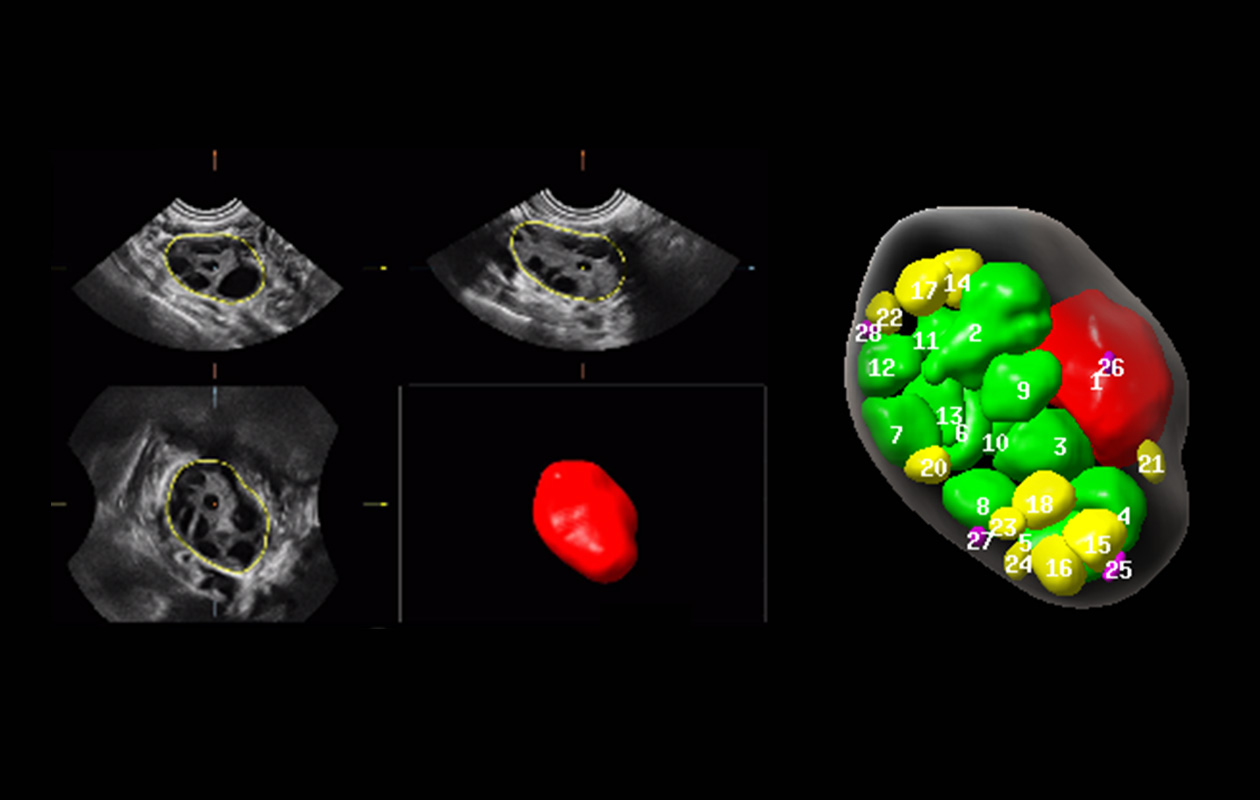
Comprehensive and intelligent volume analysis for both the ovary and follicles (3D)

Ultrasound plays a pivotal role not only in the regular examination of uterine, ovarian and adnexa, but also in the detection and characterization of abnormalities. Effective and efficient scan is enabled by a series of smart tools.
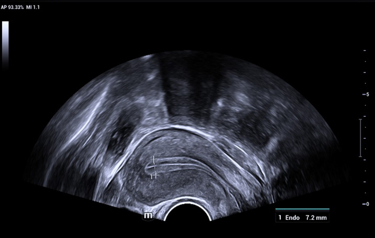
Smart Scene 2D – endometrium
Auto endometrial thickness measurement
Smart FLC -2D
Auto follicle & ovary measurements
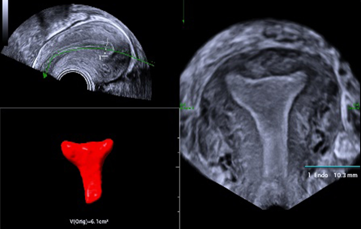
Smart ERA
Auto volume and coronal imaging
Multiple imaging modes and analysis tools allow comprehensive evaluation of the lesion, including 2D/3D tissue structure, hemodynamics, micro-flow perfusion, tissue stiffness, and quantification, giving more confidence for diagnosis, especially of complex GYN tumors.
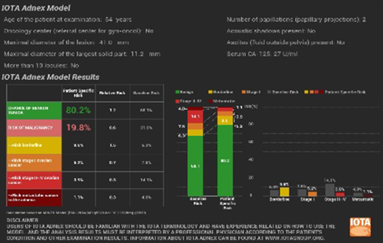
IOTA-ADNEX
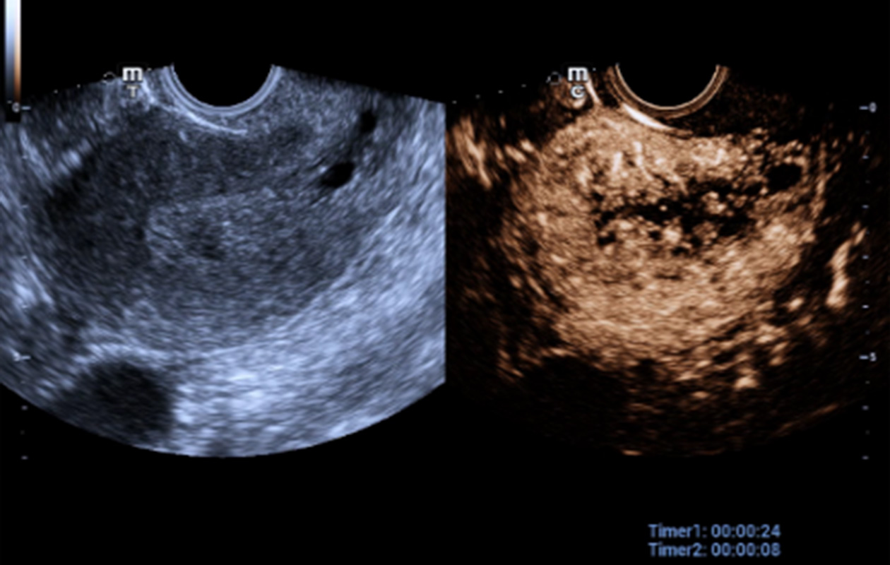
UWN+ CEUS (Cervical cancer)
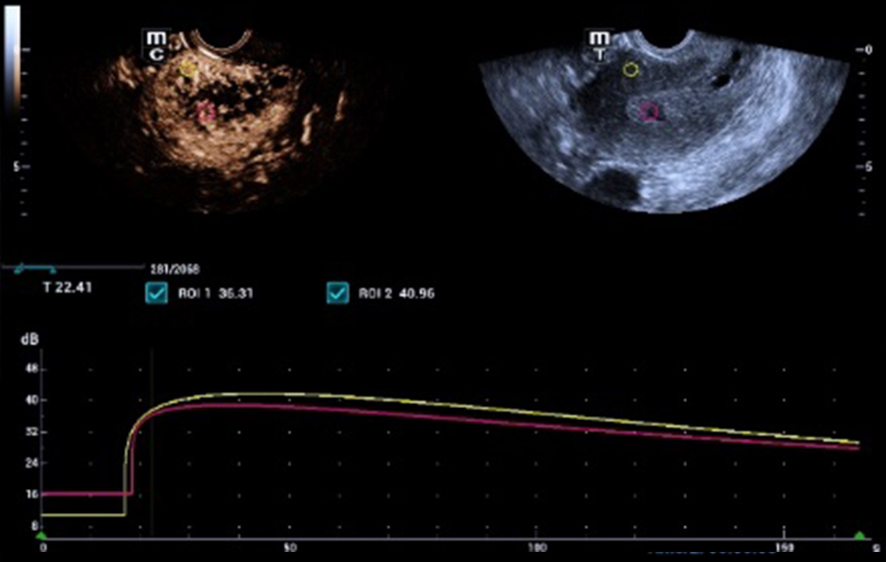
QA of CEUS
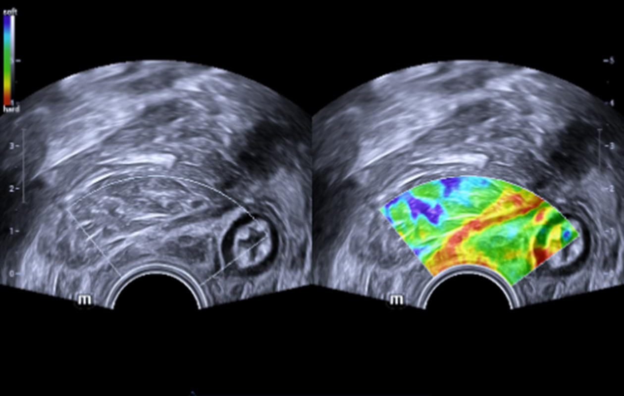
Endocavity NTE- Levator ani
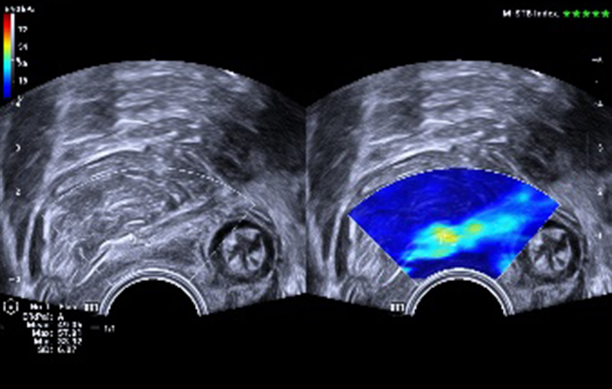
Endocavity STE - Levator ani
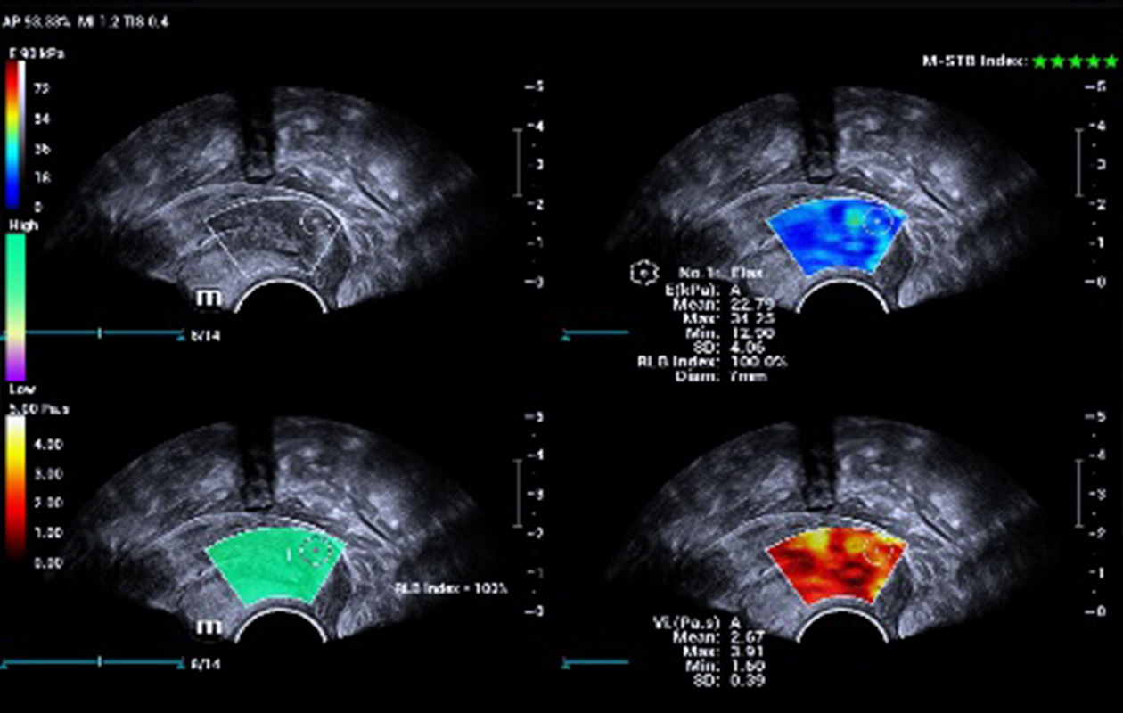
Endocavity STVi - Cervix

Accurate and standardized evaluation of dysfunctional pelvic floor, simple and ease of use, saving time up to 2/3.
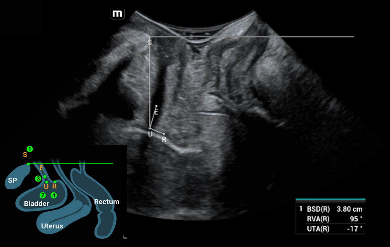
Fully automatic 2D evaluation for anterior pelvic compartment
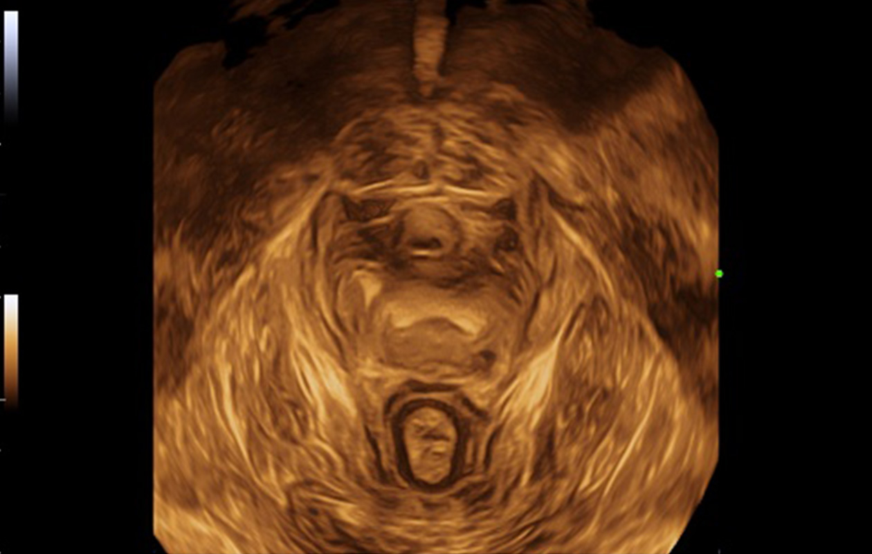
Auto volume imaging and optimization of levator hiatus
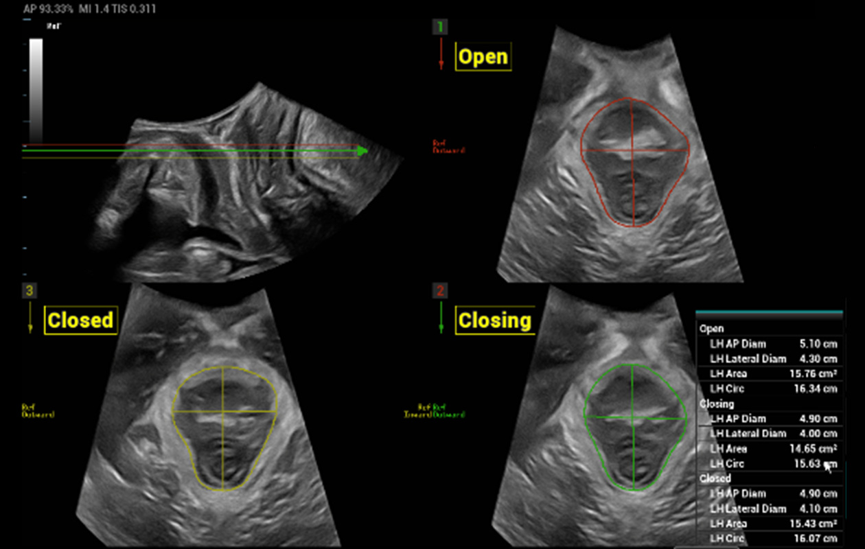
Auto planes while symphysis pubis is“open-closing-closed” and fully automatic measurements

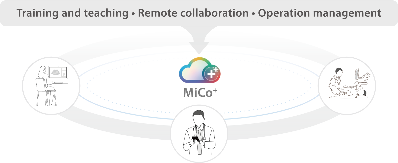
Expanding boundaries through an integrated ecosystem of devices, IT, and intelligence for elevated quality, standardized exams, and efficient management.
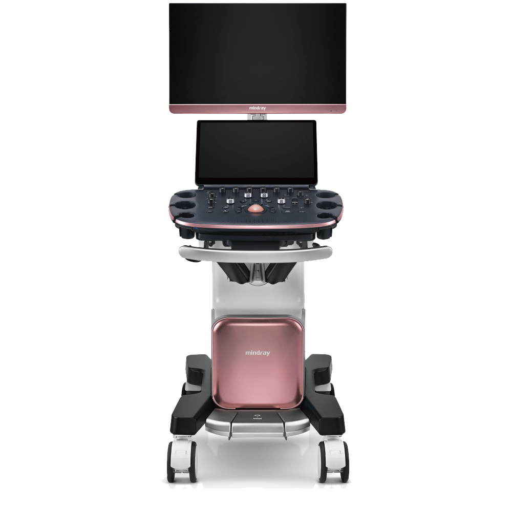
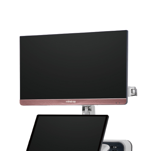
HD monitor
Full-screen view
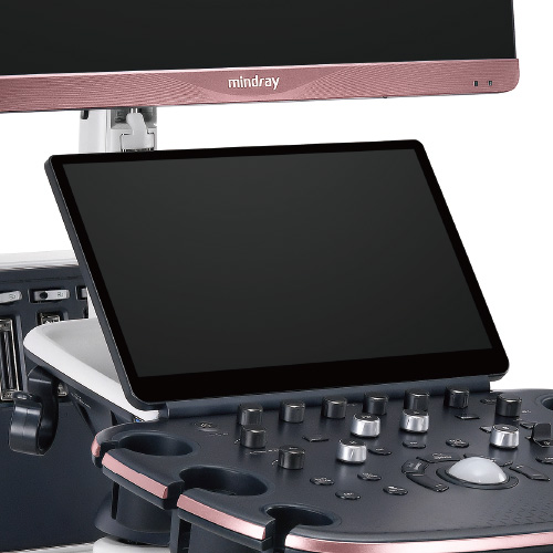
Touchscreen with
intuitive interaction
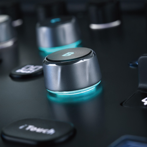
iConsole with E-ink keys
Wear-resistant buttons
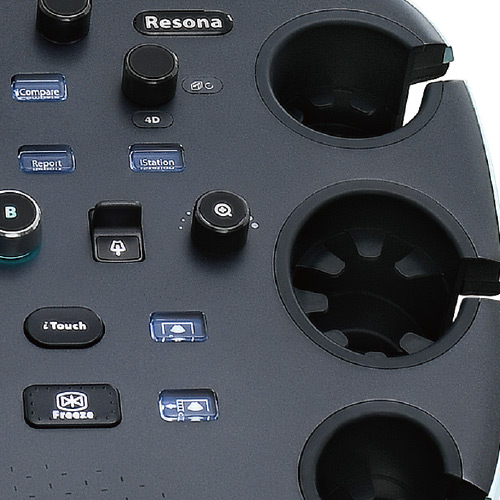
Seamless probe holders
Disinfection-friendly panel
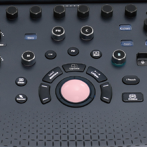
RGB illuminated trackball
Superior panel with texturing
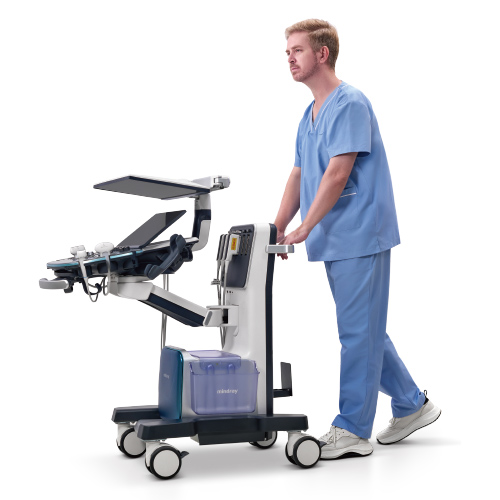
Full-space floating panel
Just fold and go with 1m height
Bedroom-level quietness
Continuous scan with battery
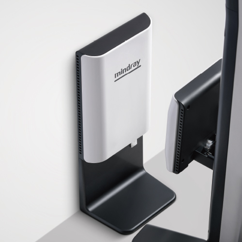
Wireless charging
Ultimate convenience