
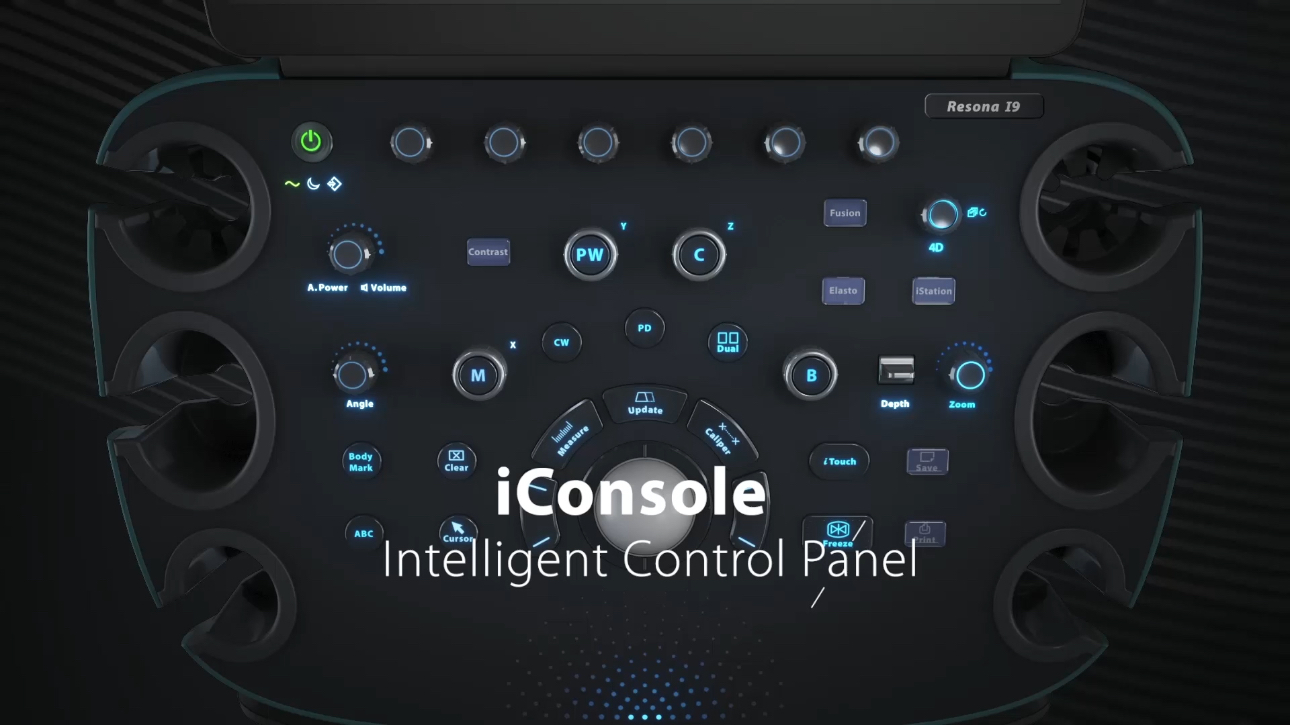
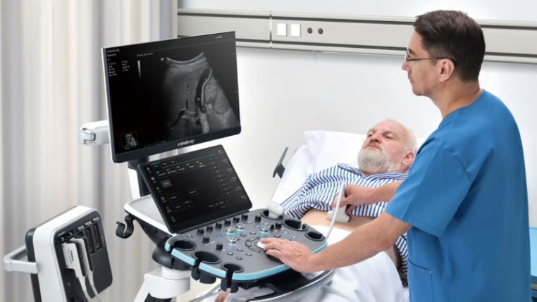
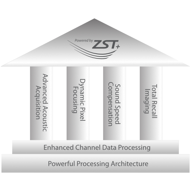
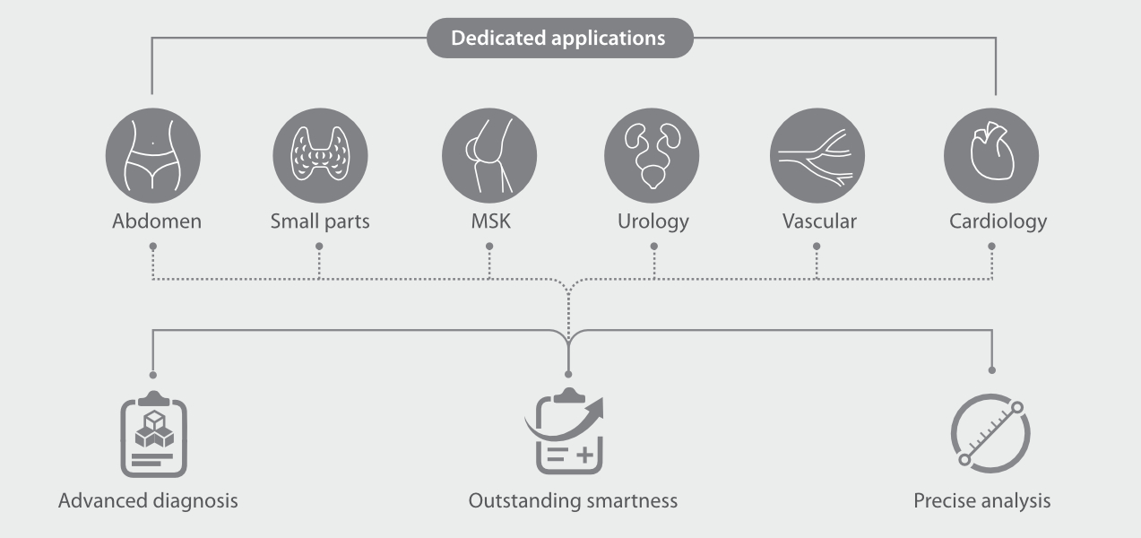
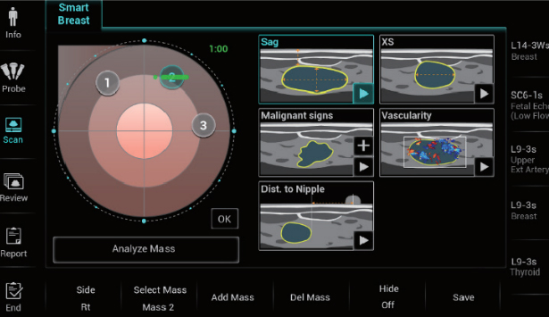
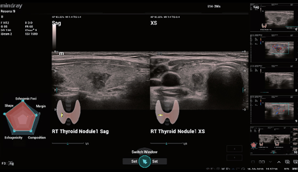
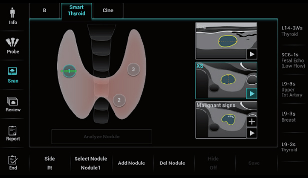
Advanced Diagnosis with Innovations
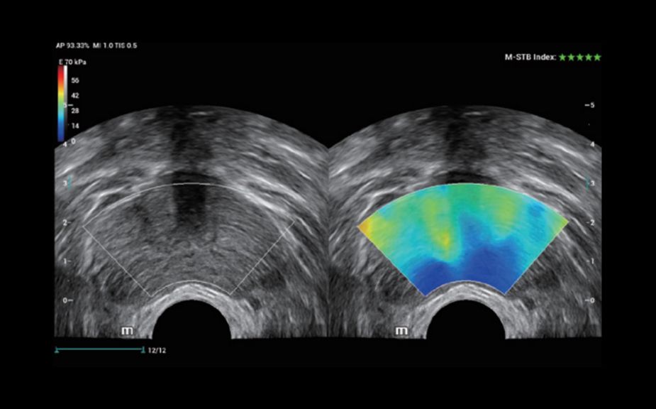
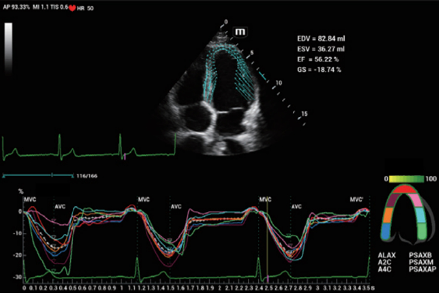
Innovative Stiffness Assessment: HiFR STE
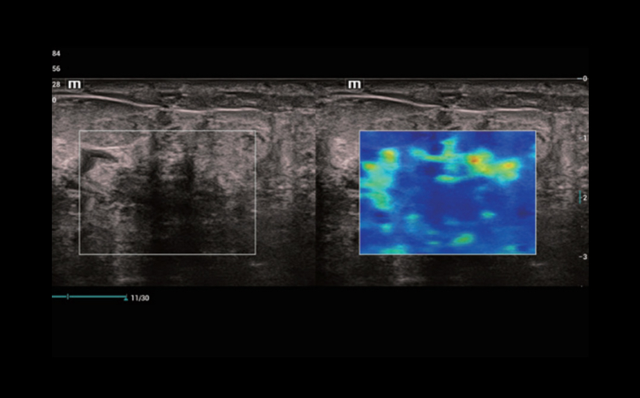
Up to 10 times faster frame rate with smooth STE imaging display
More sensitive motion detection for better stability and greater accuracy
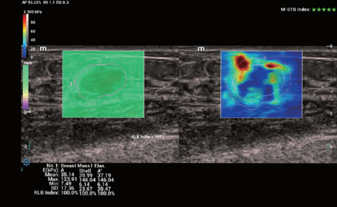
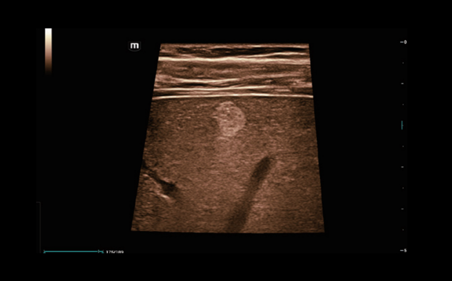
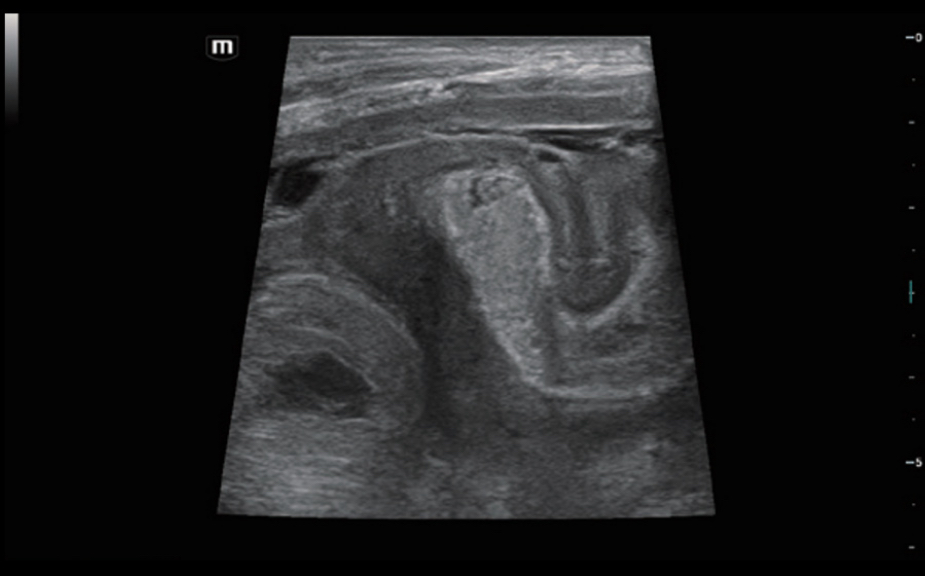
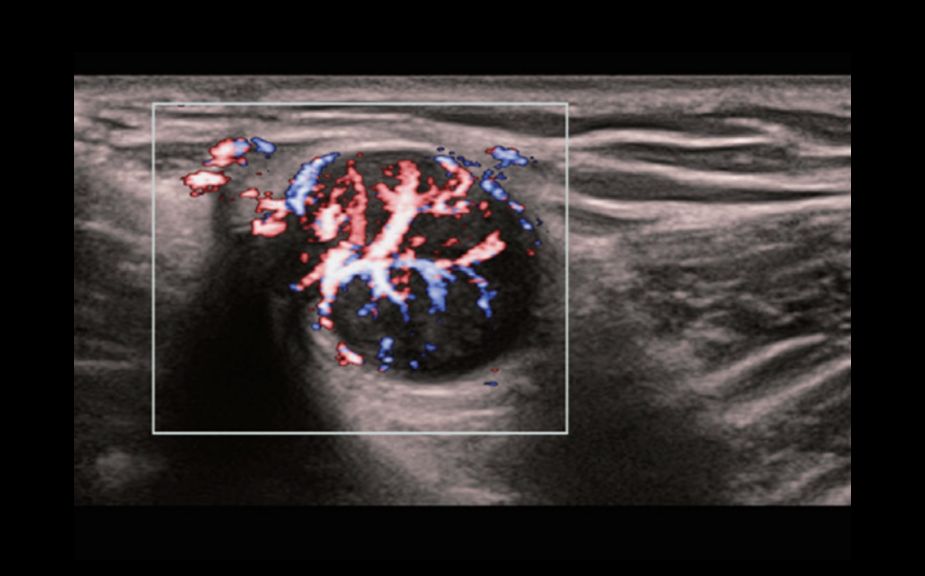
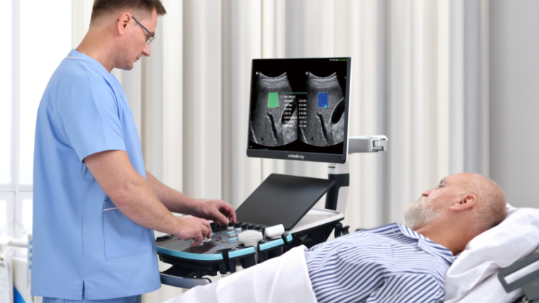
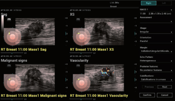
HiFR STE of breast cancer
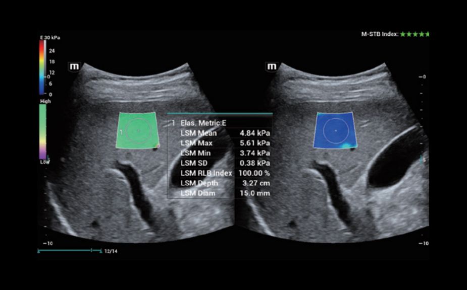
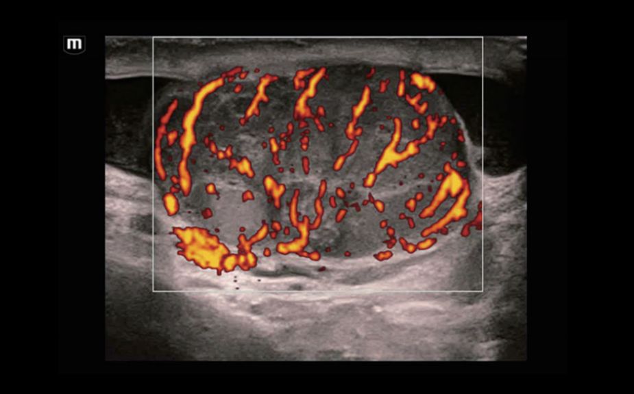
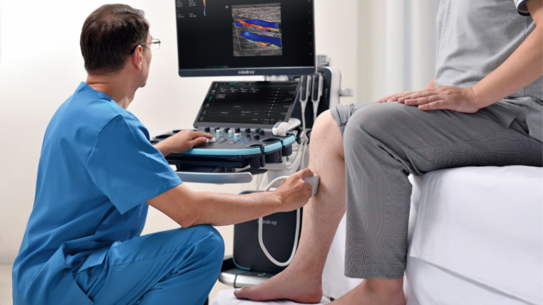
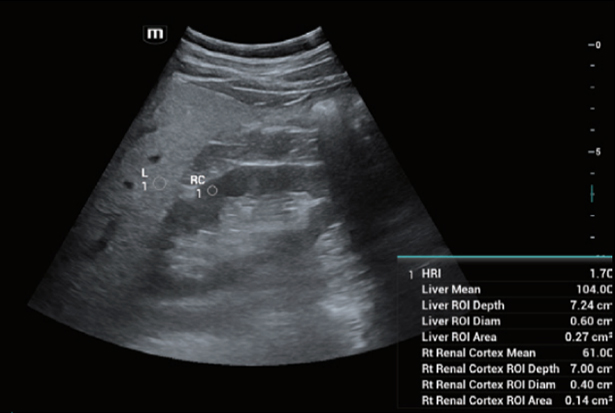
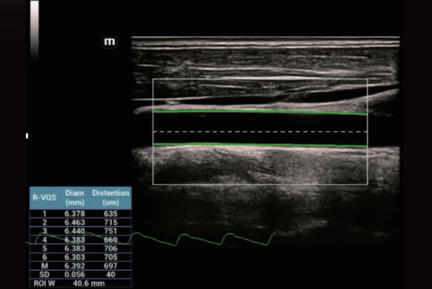
HiFR STE of liver
Innovative Stiffness Assessment: HiFR STE
Up to 10 times faster frame rate with smooth STE imaging display
More sensitive motion detection for better stability and greater accuracy
HiFR STE of breast cancer
HiFR STE of liver