Value of HiFR CEUS in Distinguishing Gallbladder Adenoma
Value of high frame rate contrast-enhanced ultrasound in distinguishing gallbladder adenoma from cholesterol polyp lesion
2023-03-14

Xiang Fei1, Nan Li1, Lianhua Zhu1, Peng Han1, Bo Jiang1, Wenbo Tang2, Maodong Sang3, Xirui Zhang3, Yukun Luo1
Department of Ultrasound1 and Department of Hepatobiliary Surgery2, The First Medical Center, Chinese PLA General Hospital, Beijing, China. Mindray Healthcare3, Shenzhen, China.
Published on European Radiology, February 2021.
Below is an issue of the Mindray Rapid Recap column - a series of article interpretation for your quick learning of the key methods and findings from the latest important research.
Background and Aim
To date, it is still a challenge to differentiate gallbladder adenomas from cholesterol polyp lesions only in the US. The vascular morphology of gallbladder polyp lesions (GPLs) detected by CEUS has been used to distinguish gallbladder adenomas from cholesterol polyp lesions.
However, for GPLs with hypervascularity, wash-in occurs rapidly and lasts only a few seconds. Therefore, a high frame rate is needed to capture these wash-in patterns. High frame rate CEUS (H-CEUS) can display microcirculation differences to increase diagnostic accuracy.
This study aimed to investigate the value of H-CEUS in distinguishing gallbladder adenomas from cholesterol polyp lesions compared to conventional CEUS and offer imaging evidence for the treatment of gallbladder adenomas and cholesterol polyp lesions.
Key Method
From July 2018 to January 2020, 94 consecutive patients with GPLs diagnosed by conventional US were finally enrolled in this study. Each patient underwent conventional CEUS and H-CEUS examinations before laparoscopic cholecystectomy. Mindray Resona 7 US system with an SC5-1U transducer was used to perform CEUS examinations (SonoVue, Bracco). Compare the perfusion features of GPLs and the final diagnosis as determined by both technologies, with histopathology as the reference.
Major Results
There were no differences in vascular types between gallbladder adenomas and cholesterol polyp lesions observed on CEUS. 84.51% of cholesterol polyp lesions and 69.57% of gallbladder adenomas were dotted vascular types observed on CEUS.
However, there were differences in vascular types between gallbladder adenomas and cholesterol polyp lesions observed on H-CEUS. 74.65% of cholesterol polyp lesions and 26.09% of gallbladder adenomas were dotted vascular types observed on H-CEUS.
- There were no differences in vascular types between gallbladder adenomas and cholesterol polyp lesions observed on CEUS. A total of 84.51% of cholesterol polyp lesions and 69.57% of gallbladder adenomas were dotted vascular types observed on CEUS.
- However, there were differences in vascular types between gallbladder adenomas and cholesterol polyp lesions observed on H-CEUS. A total of 74.65% of cholesterol polyp lesions and 26.09% of gallbladder adenomas were dotted vascular type observed on H-CEUS.
- In the cholesterol polyp lesion group, there were no differences in vascular types between CEUS and H-CEUS, while in the gallbladder adenoma group, the vascular types were significantly different between CEUS and H-CEUS.
- CEUS could detect only 4.35% of branch-like vascular type in gallbladder adenomas, while H-CEUS could detect 43.48%of branch-like vascular type in gallbladder adenomas.

-- CEUS images of a cholesterol polyp lesion show dotted vascular type (b, c).
-- H-CEUS images of a cholesterol polyp lesion show dotted vascular type (d, e, f).
The arrow indicates the vascular type of a cholesterol polyp lesion
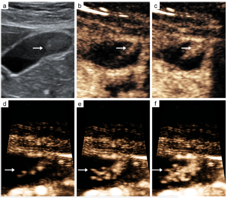
--CEUS images of a gallbladder adenoma show single vascular type (b, c).
--H-CEUS images of a Gallbladder adenoma show branch-like vascular type (d, e, f).
The arrow indicates the vascular type of a gallbladder adenoma
Discussion
The current treatment strategy recommends that follow-up is appropriate for patients with cholesterol polyp lesions, while cholecystectomy is appropriate for patients with gallbladder adenomas. Therefore, distinguishing gallbladder adenomas and cholesterol polyp lesions is important for patients with GPLs.
Interestingly, the vascular types of gallbladder adenomas were different from that of cholesterol polyp lesions observed on H-CEUS. A total of 30.43% of gallbladder adenomas were branch-like and single vascular types observed on CEUS, while 73.91% of gallbladder adenomas were branch-like and single vascular types observed on H-CEUS.
When the frame rate was increased, the time resolution of CEUS was improved to capture the perfusion progression of GPLs in detail during the early arterial phase. It is well known that the vascular types of GPLs depend on the microbubble motion trajectory in the early arterial phase. Therefore, H-CEUS could better show the details of microbubble motion trajectory compared to CEUS and thus depict the vascular morphology of a GPL more clearly and accurately.
Conclusions
H-CEUS improved the time resolution by increasing the frame rate, which helped to accurately reflect the difference in the microcirculation of GPLs and improved the ability of a differential diagnosis between cholesterol polyp lesions and adenomas. H-CUES may provide an effective means of imaging for patients with GPLs regarding the choice of treatment options.