Special thanks to Wang Jianmei, Zhang Shaojing, The Second Hospital of Tianjin Medical University.
In this journal, we will show you three cases on IUD downward displacement with septate uterus, inappropriate IUD shape, and IUD rotation, respectively.
Case 1 - IUD downward displacement with septate uterus
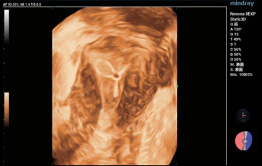
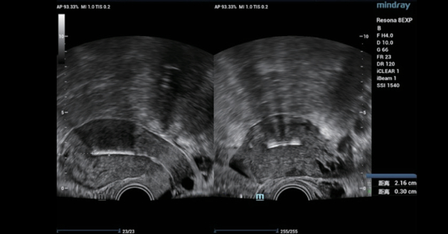
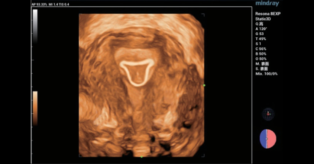
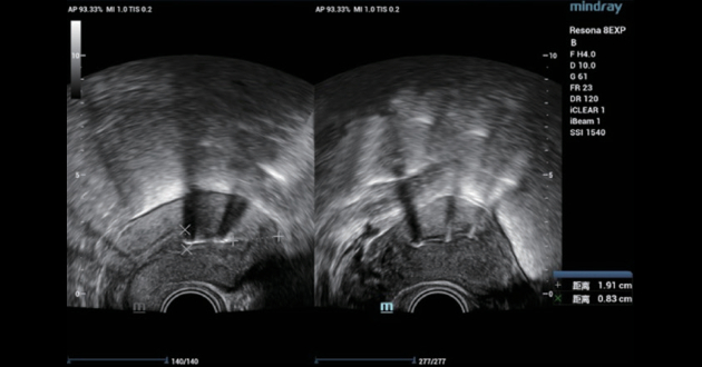
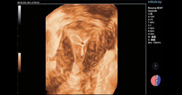
A 38-year-old female who has had an IUD insertion four years ago, diagnosed with downward displacement of IUD was admitted to our hospital for further treatment. 2D ultrasound showed mild downward displacement of the IUD. 3D ultrasound further revealed a partial septate uterus. (Figure 1 and Figure 2). The 2D ultrasound did not indicate the intrauterine morphology due to her thin endometrium.
Case 2 - Inappropriate IUD shape
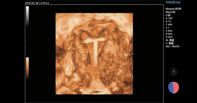
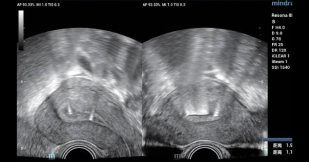
A 34-year-old female with an IUD insertion at another hospital one month ago came to our hospital for an ultrasound reexamination. The 2D ultrasound showed the IUD was at a normal position, but the 3D ultrasound showed the IUD has moved down significantly and the contraceptive effectiveness was impaired (Figure 3 and Figure 4). Therefore, we replaced the IUD with a different shape of IUD, according to the transverse diameter of the patient's uterine cavity shown by 3D ultrasound.
Case 3 - IUD rotation
A 30-year-old female, IUD insertion three months ago, with intermittent abdominal pain and abdominal distention for one month. The 2D ultrasound showed no obvious abnormality, but the 3D ultrasound suggested a rotation of the IUD (Figure 5 and Figure 6). The patient's symptoms were relieved after replacing the IUD.
Discussion
Diagnostic criteria of IUD position by 2D ultrasound
・IUD in Normal Position
On the maximum longitudinal view of the uterus, the distance between the upper edge of the IUD and the perimetrium of the uterine fundus should be ≤ 2 cm (acceptable range: 1.7–2 cm).
・Ectopic IUD
- Downward displacement: On the maximum longitudinal view of the uterus, the distance between the upper edge of IUD and the perimetrium of the uterine fundus is > 2 cm;
- IUD incarceration: The IUD is fully or partially implanted into the myometrium;
- IUD perforation: The IUD fully or partially penetrates through the perimetrium into the pelvic cavity or abdominal cavity.
Advantages of 3D Ultrasound
The 3D ultrasound can clearly judge the position relation between the IUD and endometrium and myometrium, further clarify the spatial position and shape of the IUD, and provide more accurate diagnostic information for the IUD incarceration and rotation. In addition, it can provide more information about the patient's uterine cavity morphology, which is helpful to select an appropriate IUD based on the shape and size of the uterine cavity. In particular, it is of great reference significance to explore the causes of various discomfort causes after IUD insertion. Therefore, 3D ultrasound has potential and extensive clinical value in the field of gynecology, especially in birth control.

