Hemabook Chapter 18: PLT-H: A new parameter for accurate platelet counting with anti-interference ability
2022-08-11

Hemostasis and coagulation are the main functions of platelets (PLTs). Thrombocytopenia is a common cause of bleeding. A PLT count from 20 to 50×109/L may indicate mild or surgical bleeding. If it is lower than 20×109/L, this may indicate severe bleeding. If it reduces to 5×10⁹/L or lower, the patient may be experiencing a life-threatening condition.
Nowadays, laboratories usually count PLTs by using automatic hematology analyzers, which work based on various methods with different characteristics.
Conventional impedance methods (PLT-I)
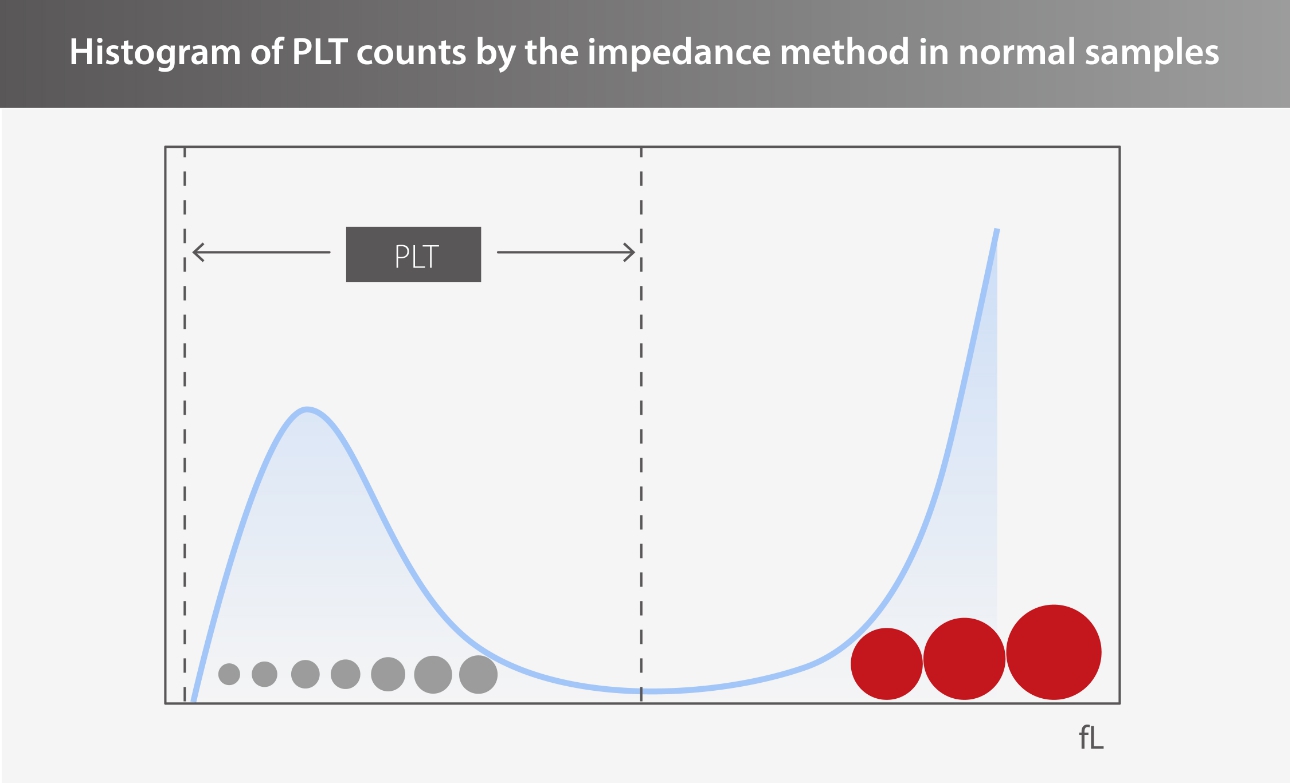
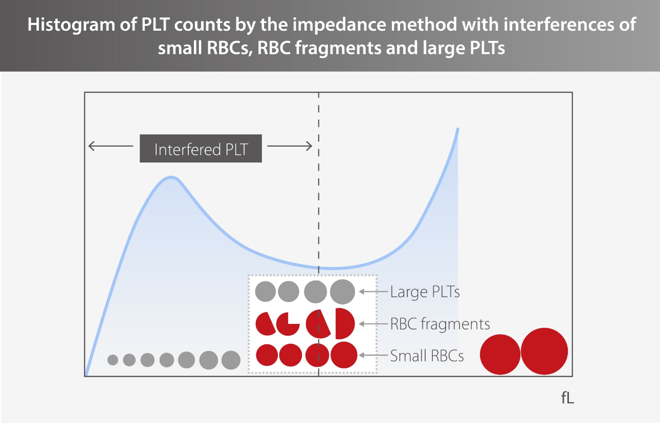
PLTs and red blood cells (RBCs) can be distinguished by the magnitude of the electrical impedance signal in normal samples. However, PLT-I is subject to the interference of microcytic RBCs, fragments, and large PLTs when it is used to differentiate PLTs by cell volume.
When microcytic RBCs and fragments are present in the blood, the result of PLT-I will be falsely high due to the interference of RBCs; when large PLTs or PLT aggregations are present in the blood, the PLT measurement result will be falsely low.
Although the PLT-O (optical platelet) method can be used to avoid the above inferences, it requires additional reagents for the analysis. PLT-H is a new parameter provided by Mindray BC-700 Series that can resist interferences in conventional PLT detection and requires no extra reagents in every CBC and DIFF analysis. Let’s take a look at how PLT-H provides accurate PLT measurement results.
Clinical case study 1
A patient with acute lymphoblastic leukemia, hospitalized for 11 months, was undergoing chemotherapy and preparing for bone marrow transplantation.
Microscopic examination revealed evident RBC fragments (arrows) in multiple high-power lens.
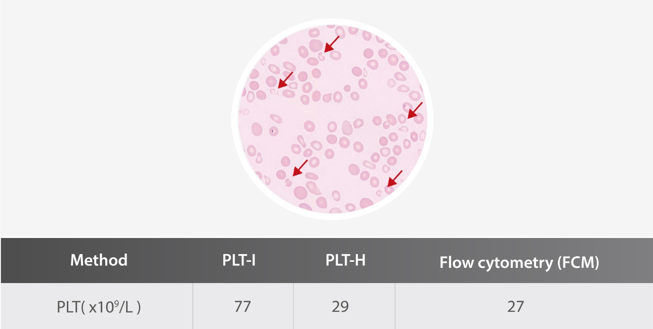
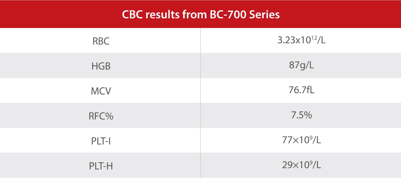
The PLT count measured by flow cytometry was 27X109/L, which was consistent with that by PLT-H.
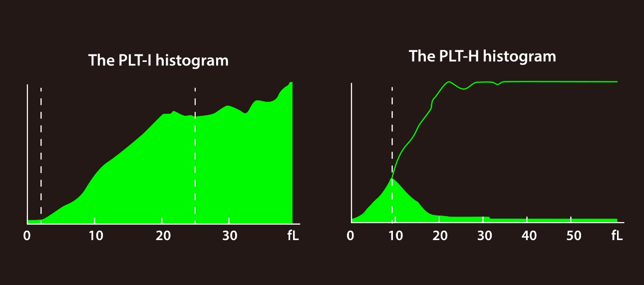
In this case, an RBC fragments alarm was triggered in the instrument, and the FRC value was 7.5%. The tail of the PLT histogram was elevated, suggesting that RBC fragments may be present in this sample, which would cause a false high value in PLT. During chemotherapy, patients usually exhibit a decrease in whole blood cells and an increase in cell fragility. Despite the presence of RBC fragments, PLT-H can accurately measure the PLT count and monitor the risk of bleeding in a timely manner.
Clinical case study 2
A patient with systematic lupus erythematosus revisited the hospital and got checked by Mindray BC-700 Series hematology analyzer.
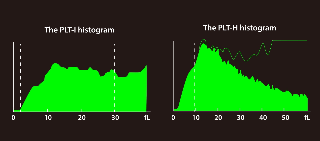
Microscopic examination revealed evident large PLTs (arrows) having a similar size to the RBCs in multiple high-power lens.
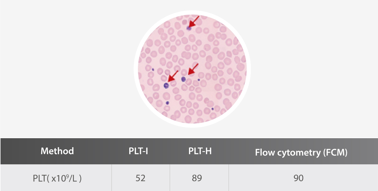
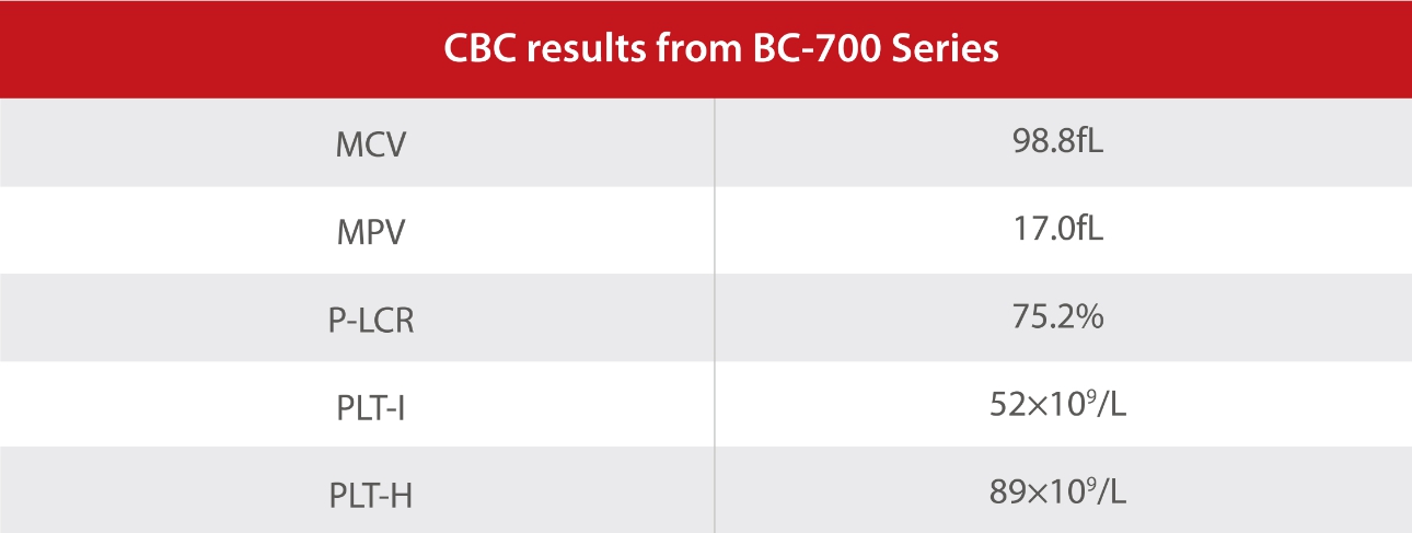
The PLT count measured by flow cytometry was 90X109/L, which was consistent with that by PLT-H.
In this case, the MPV and P-LCR values increased significantly. The tail of the PLT histogram was elevated, suggesting that large PLTs may be present in this sample, which would cause a pseudo-thrombocytopenia. The information obtained from the DIFF channel can ensure the accurate detection of large PLTs, avoiding unnecessary blood transfusion.
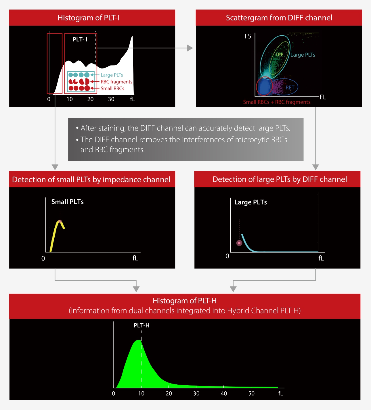
Principle of brand-new parameter - PLT-H
From the impedance channel, the large PLTs are interfered by microcytic RBCs and fragments, but the small ones are not. From the DIFF channel, the RBCs are dissolved by specific reagents, the PLT structure remains intact, and large PLTs are detected by the precise optical methodology. By combining the small PLTs from the conventional impedance method and the large ones from the optical method, an accurate PLT count will be obtained.
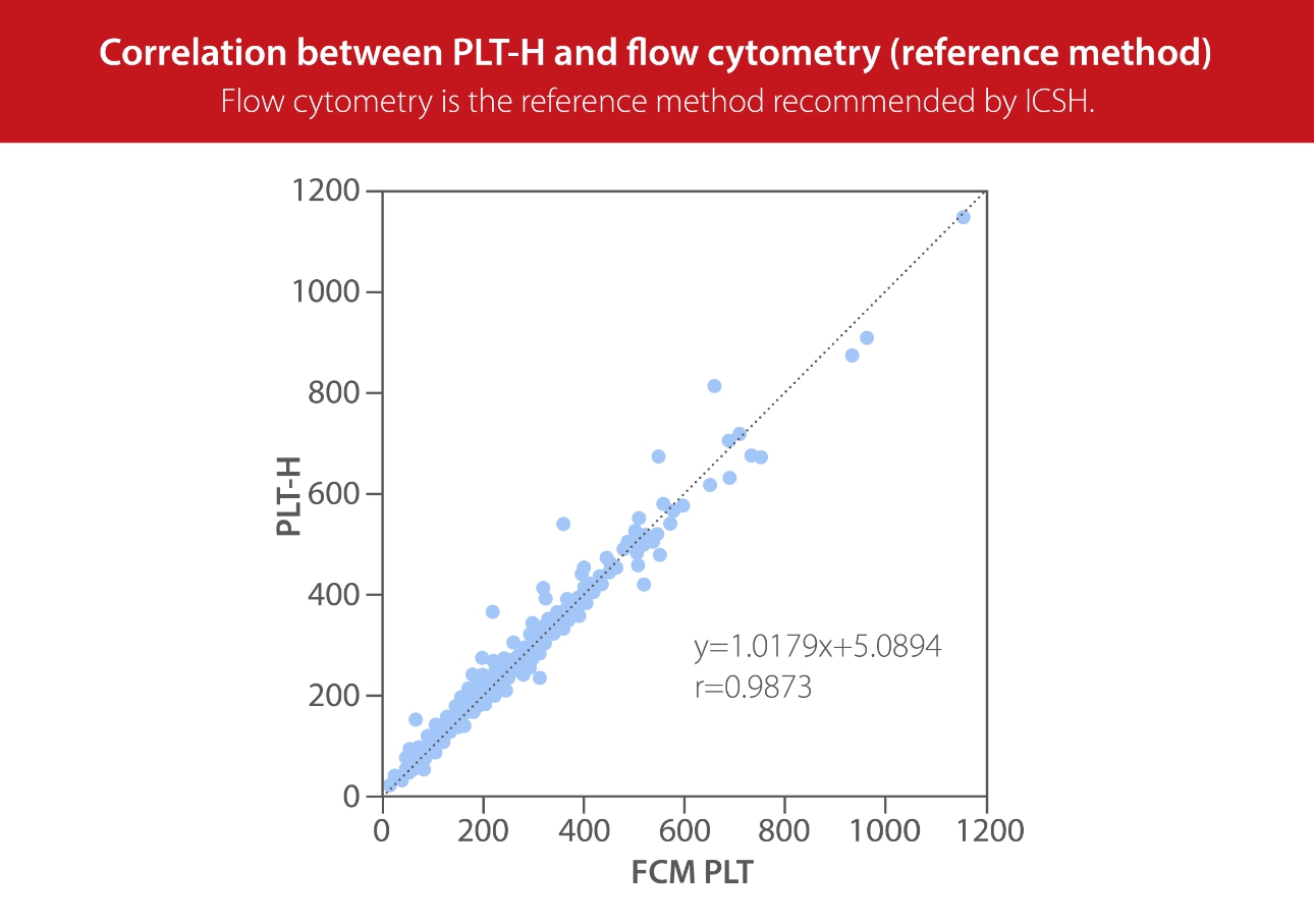
In Thailand, 405 PLT samples were collected(excluding aggregation samples). As revealed by microscopic examination, 200 samples contained interfering factors, including large PLTs, microcytic RBCs and RBC fragments. The PLTs were detected by Mindray BC-760 and BD flow cytometers, respectively. BC-760 PLT-H had a good correlation with flow cytometry (r=0.9873, y=1.0179x+5.0894).
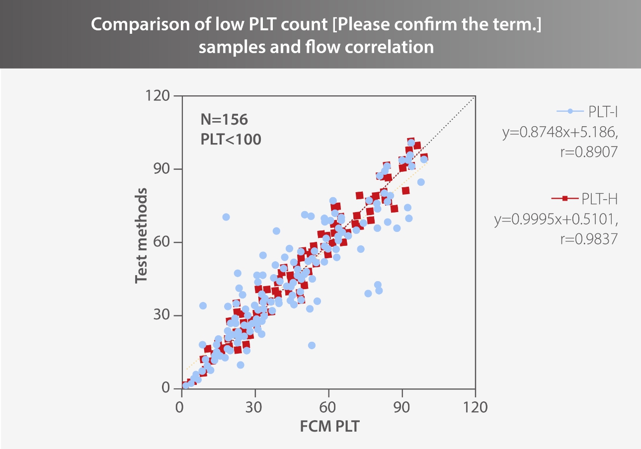
156 low PLT count [Please confrm the correct term.] samples were selected. Using flow detection (CD41+CD61) as the reference method, the correlation between PLT-H and PLT-I in the low-count [Please confirm the correct term.] segment of Mindray’s hematology analyzer was analyzed, as shown in the figure. It can be seen from that PLT-H has a strong correlation with flow cytometry (r=0.9837, y=0.9995x+0.5101).
In conclusion, the use of PLT-H technology can significantly reduce the inaccurate counts caused by the interferences of RBC fragments, large PLTs, and small RBCs, helping greatly in generating accurate and reliable PLT detection reports for clinical purposes.