Ultrasound Journal 12 - Multiparametric Ultrasound in Differentiating a Complex Cystic Renal Mass: A Case Report
2023-03-21

Introduction
A 52 years old man was found with a cystic lesion in the right kidney when he underwent an abdominal ultrasound scanning for preventive examination. A preliminary diagnosis of an indeterminate renal cystic mass was made. Then he was advised to do further tests for characterization of the cystic renal mass.
Ultrasound examination
1) B-mode and Power Doppler mode imaging
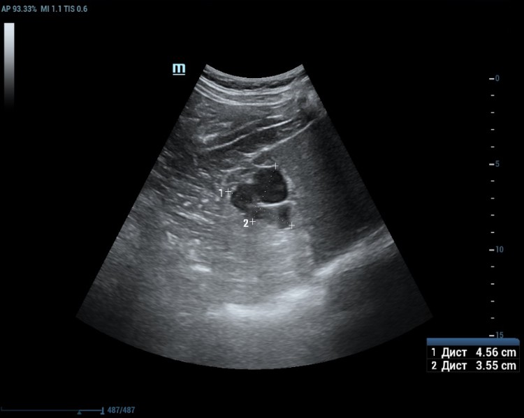
An ultrasound examination was conducted using a Resona 7 ultrasound machine (Mindray, China) with the SC5-1U transducer. A cystic lesion measuring 46x35 mm in the middle part of the right kidney was found with multiple septa measuring up to 2 mm thick. When scanning the right kidney, a cystic mass with clear, even contours, 46 x 35 mm in size with multiple thick partitions (about 4 partitions) is visualized in the middle part of the cortex. It is homogeneous and anechoic within the cystic mass. A hyperechoic node measuring 10 mm in length was seen protruding into one of the cystic partition (Figure 1 and 2).

Power Doppler was used to evaluate for internal flow within the complex cystic mass with indeterminate results. (Figure 3).

2) Contrast Enhanced Ultrasound (CEUS)
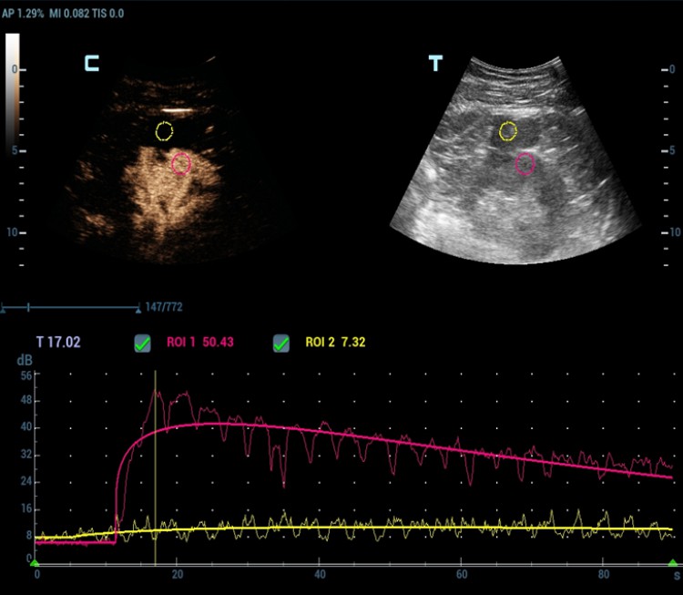
As Power Doppler showed indeterminate results regarding internal flow within the complex cystic mass, CEUS was performed through injection 1.2 ml of Sonovue (Bracco Swiss, SA, Switzerland). The longitudinal plane of the kidney with the maximum section of the cystic mass was selected for scanning. At the end, the cystic mass was characterized as benign by a lack of enhancement throughout the CEUS study (Figure 4 and 5).


Quantitative analysis also confirmed the absence of enhancement within the cystic mass (Figure 6).
Сomputed tomography (CT)
The patient was also performed an abdominal CT examination with intravenous contrast of 50ml Optirei 350. The CT study demonstrated a multilocular parenchymal cyst with a size of 40x40 mm in the middle of the right renal cortex. Subcapsular bulging beyond the renal cortex with a small accumulation of contrast and the presence of small calcifications in the cyst wall was revealed. CT examination indicated that it was a multilocular complex cyst of the right kidney.
Pathology result
Despite the fact that there were no signs of malignancy for the renal cystic mass, the patient insisted on surgery. The cystic mass of the right kidney was resected at the Republican Clinical Oncological Dispensary of the Ministry of Health of the Republic of Tatarstan named after Professor M.Z. Sigal. Finally, the histological examination confirmed a simple renal cyst (Figure 7).

Discussion
Ultrasound examination is the most common method for visualizing the kidneys. With B-mode ultrasound, usually it is easy to differentiate a simple cystic mass. But sometimes it is difficult to differentiate complex cysts from malignancy, even with Doppler imaging. However, the introduction of ultrasound contrast agents into clinical practice has significantly improved the diagnostic efficiency of ultrasound. CEUS is very useful in the further assessment of uncertain cystic renal mass as it can show whether there is enhancement or not [1,2,3]. In this study, power Doppler was used to evaluate for internal flow with indeterminate results. But CEUS demonstrated that there was no enhancement within the complex cystic mass and the mass was characterized as benign. Finally, the postoperative histological examination confirmed the same diagnosis. Therefore, CEUS is very helpful for non-invasive characterization of complex cystic renal mass and may help patients avoid surgery.
A special thanks for sharing this case to PhD Dr. Hasanov Marat Zufarovich from the Ultrasound Department of the Republican Clinical Oncological Dispensary of the Ministry of Health of the Republic of Tatarstan named after Professor M. Z. Sigal.
Reference
[1] Sidhu PS, Cantisani V, Dietrich CF. et al. The EFSUMB Guidelines and Recommendations for the Clinical Practice of Contrast-Enhanced Ultrasound (CEUS) in Non-Hepatic Applications: Update 2017 (Short Version). Ultraschall in Med 2018; 39: 154-180
[2] Barr RG, Peterson C, Hindi A. Evaluation of indeterminate renal massess with contras-enhanced US: a diagnostic performance study. Radiology 2014; 271: 133-142
[3] Rubenthaler J, Bogner F, Reiser M. et al. Contrast-Enhanced Ultrasound (CEUS) of the Kidneys by Using the Bosniak Classification. Ultraschall in Med 2017; 37: 234-251