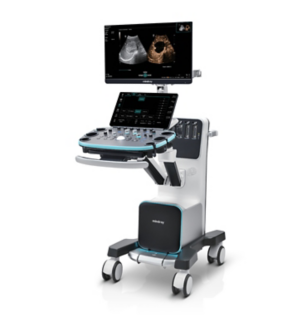
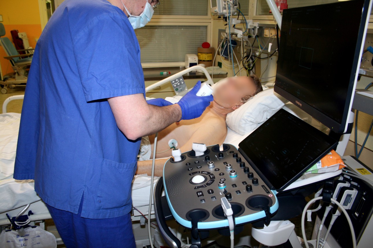
When working in an intensive care unit, obtaining a quick overview of a patient's health status is crucial. Sonography can non-invasively image multiple entities, some of which may be critical, in real-time. However, using this technology requires equipment that offers high flexibility without compromising image quality. For his department, specialized intensive care physician Dr. Armin Seibel relies on the high-end color Doppler ultrasound device Resona I9 from Mindray, which offers exceptional image quality, multifunctionality, and a long battery life.
Kirchen, a small town in Rhineland-Palatinate nestled among wooded low mountains, is home to the local hospital, which has just over 270 beds. As the only medical center in the rural area, it provides basic and standard medical care as well as emergency services for the community. Dr. Armin Seibel, a specialist in anesthesiology, intensive care medicine, and emergency medicine, manages the department in an interdisciplinary style. He treats a diverse range of patients with various clinical conditions, requiring multi-organ ultrasound diagnostics expertise.


Diverse application possibilities
Dr. Seibel chose Resona I9 for various reasons. First, the high-end device offers excellent image quality with high resolution, as well as focused examination techniques that facilitate diagnosis. For instance, he frequently uses contrast-enhanced ultrasound (CEUS) to clarify uncertain findings.
Versatility and Long Battery Life Matters
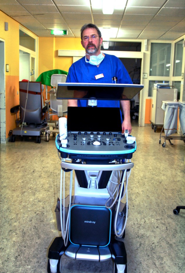
Resona I9 provides all the tools Dr. Seibel needs for widespread use in the intensive care unit. In addition to versatile application possibilities, another critical advantage is its long battery life of up to four hours. It can accompany a complete morning round without the need for frantic searches for power outlets. Furthermore, it is challenging to locate free sockets near an intensive care patient because of numerous connected machines. "An ultrasound device that requires plugging into a socket is rarely unplugged to reach the other side of the patient. Instead, it is customary to pull the probe and cable over the patient. However, I consider this practice unsanitary, especially in the ICU," explains Dr. Seibel. A high battery capacity ensures continuous wireless operation and contributes greatly to convenience and infection control.
About Dr. Armin Seibel
Dr. Armin Seibel is an accomplished specialist in anesthesiology with additional qualifications in special intensive care medicine and emergency medicine. Currently serving as a senior physician for interdisciplinary intensive care medicine at German Red Cross Hospital Kirchen, Dr. Seibel has established himself as an expert in emergency sonography. He has been a DEGUM-certified course instructor Level III since 2014 and serves as a speaker for DEGUM Working Group on Emergency Sonography. In addition to his clinical work, Dr. Seibel is also involved in medical education and clinical research. He is passionate about firefighting medicine and actively serves as a firefighter physician.