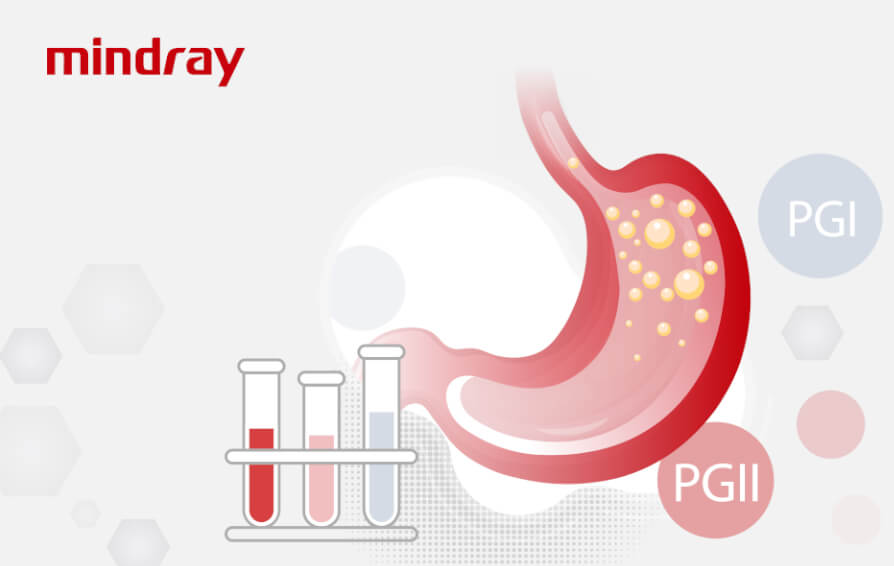
Pepsinogen is a powerful and abundant protein digestive enzyme. It is synthesized and secreted primarily by the gastric chief cells as a zymogen. Pepsinogen is then converted by gastric acid in the gastric gland into the active enzyme pepsin, which is crucial for digestive processes in the human stomach. Also, pepsin can activate additional pepsinogen autocatalytically.
Pepsinogen physiology
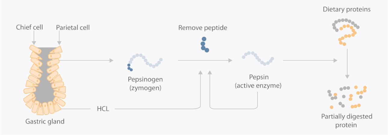
Pepsinogen has two subtypes: pepsinogen I(PGI) and pepsinogen II(PGII).
Gastric PG I and II are produced by the chief cells in the gastric fundus. PG II is also produced by the pyloric glands in the antrum and Brunner’s glands.

Serum PGI and PGII

Pepsinogen is present in gastric mucosa. A small amount of pepsinogen may be released into the blood. When pathological changes occur in the gastric mucosa, the serum pepsinogen level changes correspondingly, reflecting the condition and function of the gastric mucosa.
Gastric cancer overview

According to recent updates to the global cancer burden statistics, the incidence rate for gastric cancer puts it in the top five, and it is one of the leading causes of death by cancer.
It is also considered one of the biggest obstacles to increasing life expectancy for both sexes worldwide. [1]
Cause of Gastric cancer
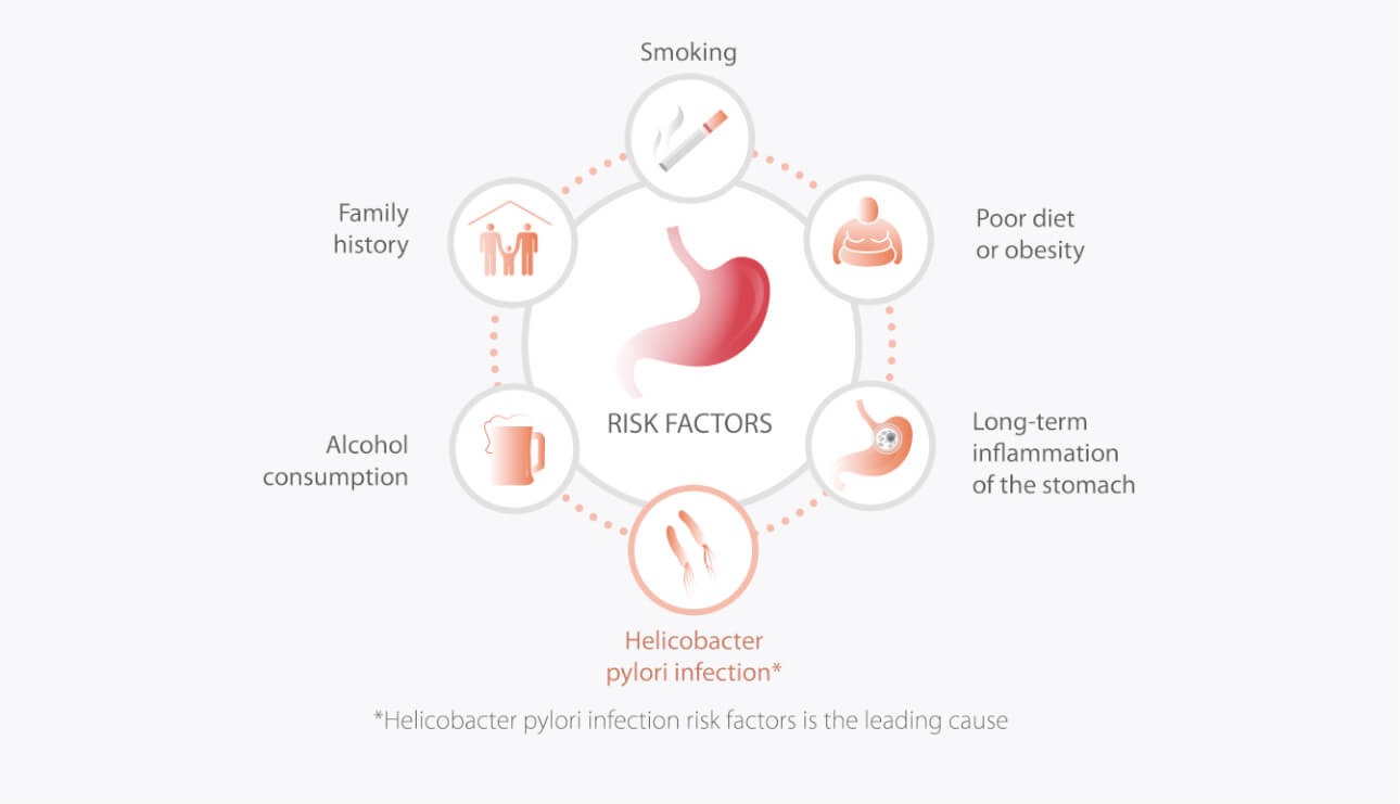
The prevalence of H. pylori infection is extraordinarily high, infecting 50% of the world’s population, and its geographic variation correlates reasonably with that of gastric cancer incidence rates.
Gastroscopy is the gold standard for chronic atrophic gastritis diagnosis. However, the invasive and painful nature of the procedure and its limited resources often restrict its clinical application. For these reasons, screening for chronic atrophic gastritis is of the utmost importance.
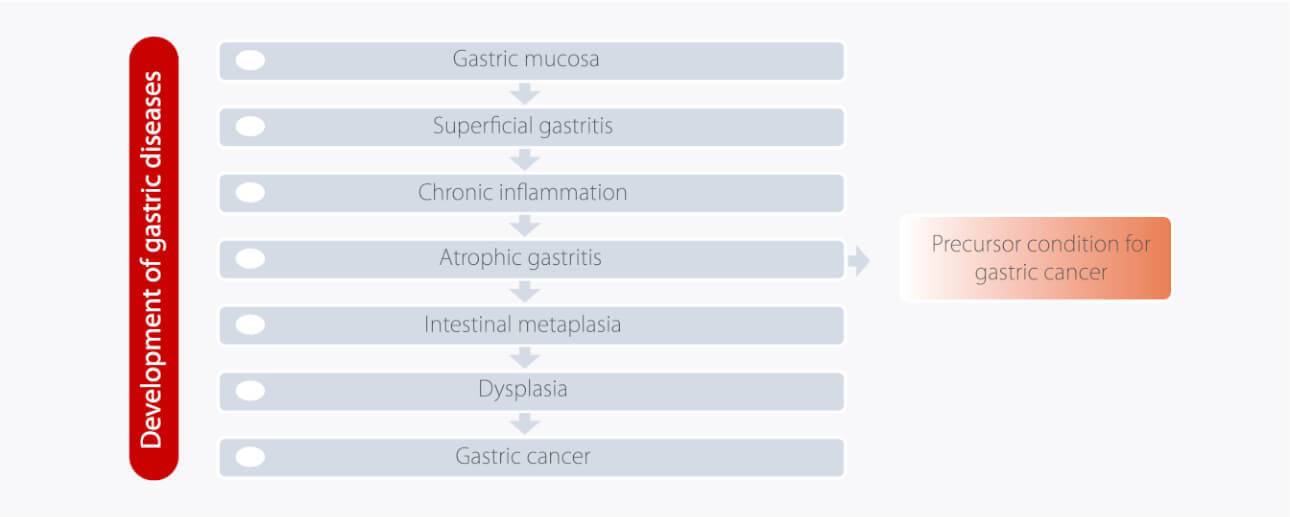
Many studies show evidence supporting the theory that serum levels of PGI and/or PGI/II ratio correlate with morphologic and functional changes in the gastric mucosa. Therefore, pepsinogens have been widely used as ‘serological biopsy’ for the screening of gastric precancerous lesions. Some guidelines on gastric cancer prevention recommend serum pepsinogen in screening for chronic atrophic gastritis. [2,3]

Clinical application

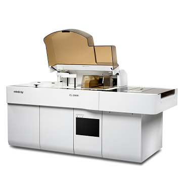
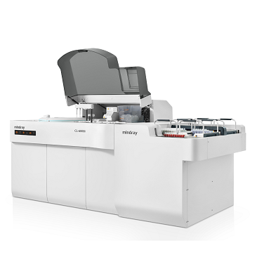
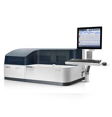
Mindray provides reliable PGI and PGII assays to screen atrophic gastric, detect H. pylori (HP) infections, well as monitor the changes after HP eradication and recurrence after total gastrectomy.

References
[1] Sung, H., et al., Global cancer statistics 2020: GLOBOCAN estimates of incidence and mortality worldwide for 36 cancers in 185 countries. CA Cancer J Clin, 2021.
[2] Lahner, E., et al., Chronic atrophic gastritis: Natural history, diagnosis and therapeutic management. A position paper by the Italian Society of Hospital Gastroenterologists and Digestive Endoscopists [AIGO], the Italian Society of Digestive Endoscopy [SIED], the Italian Society of Gastroenterology [SIGE], and the Italian Society of Internal Medicine [SIMI]. Dig Liver Dis, 2019. 51(12): p. 1621-1632.
[3] Fock, K.M., et al., Asia-Pacific consensus guidelines on gastric cancer prevention. J Gastroenterol Hepatol, 2008. 23(3): p. 351-65.
[4] 蒋孟军,肖志坚.胃癌患者血清胃蛋白酶原含量的检测及其临床意义[J].实用癌症杂志,2000(01):40-42.
[5] Hiroki, O. , et al. "A significant increase in the pepsinogen I/II ratio is a reliable biomarker for successful Helicobacter pylori eradication." Plos One 12.8(2017):e0183980.
[6] Mansour-Ghanaei, F. , et al. "Only serum pepsinogen I and pepsinogen I/II ratio are specific and sensitive biomarkers for screening of gastric cancer." Biomolecular concepts 10.1(2019):82-90.
[7] Massarrat, S. , et al. "Pepsinogen II Can Be a Potential Surrogate Marker of Morphological Changes in Corpus before and after H. pylori Eradication." Biomed Research International 2014(2014):481607.
[8] Ping Li, et al. “Pepsinogen I and II expressions in situ and their correlations with serum pesignogen levels in gastric cancer and its precancerous disease.” BMC Clinical Pathology 2013, 13:2.
