Introduction
This case highlights endometrioma as an unexpected etiology of a vaginal cyst. The diagnosis of endometriosis is surprising when it is found outside the walls of the peritoneal cavity and is challenging without histology confirmation. Endometriosis can “masquerade” as several other conditions, often leading to missed or delayed diagnosis.
Mechanism
Few cases of endometriosis in the vagina have been described. Review of the literature demonstrated endometriosis in an episiotomy scar with a tender nodule and cyclic pain. Similar to the patient described here, another report revealed a vaginal endometrioma in a 43-year-old patient who had no other symptoms of endometriosis. Clinical impression preoperatively was most consistent with urethral diverticulum, whereas MR and US imaging suggested Gartner’s duct cyst. She remained asymptomatic three months postoperatively.
A 26 years old patient, G0P0, complained of a feeling of a foreign body in the perineum during long walking, physical exertion, soreness during sexual intercourse, as well as copious painful menstruation with menarche. The patient discovered the formationin the perineum area 8 months ago, according to the words, it was slowly increasing.
From anamnesis: Menstrual cycle from the age of 14, regular, after 28-29 days, 7-9 days, abundant and very painful (always takes NSAIDs).
Objectively examined: Height 172 cm, weight 57 kg, BMI 19 kg/m2
Ultrasound of the pelvic organs: signs of the proliferation phase, diffuse form of adenomyosis

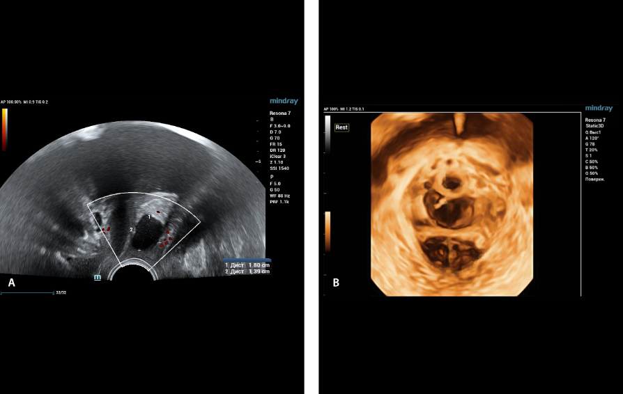
During ultrasound examination using Endocavity volume convex array transducer (DE10-3WU, Resona 7, Mindray) in the projection of the anterior vaginal wall along the middle third of the urethra, an ovoid-shaped formation with dimensions of 18x14 mm with a parietal fine suspension, non-displaced by transducer traction, sensitive, with single vascular loci in the CDI mode is visualized. In 3D reconstruction – formation with heterogeneous contents, with hyperechoic septa.
Treatment plan
This patient was referred to surgeons for the surgical removal of this formation (cystectomy) and preservation of the formation capsule, as well as hysteroscopy and endometrial biopsy, taking into account the patient's complaints. In the postoperative period, long-term therapy with progestogens (dienogest 2 mg) for at least 6 months is recommended.
Conclusion
Volumetric reconstruction of pelvic floor formations makes it possible to clarify the diagnosis and correctly choose the method of treatment, and also serves as a navigational guide for the surgeon.
