


Special thanks to Dr. Huang Pintong and Lin Tao, The Second Affiliated Hospital of Zhejiang University School of Medicine.
Key points:
- Primary Pancreatic Lymphoma (PPL) is a rare malignant tumor which is difficult to diagnose
- CEUS appearance could be an important indicator for suspicion of PPL
Case Description:
A 64 year-old male presented to his local hospital with acute left upper quadrant pain where B-mode ultrasound demonstrated "heterogeneous hypoechogenicity in the splenic area and splenic space-occupying lesions". He was then transferred to a higher-level hospital where CT and MRI showed space occupation in the pancreatic tail, spleen, and left perirenal space. His routine blood cell count, liver, kidney functions and tumor marker tests were all normal. He was then referred to the Second Affiliated Hospital of Zhejiang University School of Medicine for final diagnosis.
Imaging Findings:
The patient underwent conventional US, CEUS, CT, and whole body PET/CT imaging with the findings presented in Fig 1-3.


Diagnosis:
All imaging reports indicated malignancy but were nonspecific for primary pancreatic cancer. An ultrasound guided biopsy was performed of the pancreatic mass with a resultant diagnosis from pathology of Burkitt lymphoma which is a primary pancreatic lymphoma.
Treatment:
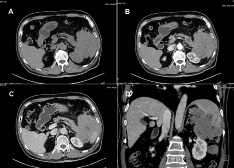
The patient underwent R-hyper-CVAD-A chemotherapy under the Hematology Department with serial CT demonstrating obvious reduction in tumor size (Fig 4 A-C). The patient also experienced a significant improvement in general overall condition.

Conclusion:
Primary pancreatic lymphoma is a very rare non-epithelial malignant tumor of the pancreas, representing only 0.5% of all pancreatic masses and only 2% of all extranodal lymphomas.[1] The clinical manifestations and laboratory tests are non-specific, and the imaging manifestations are easily confused with other pancreatic malignancies such as pancreatic ductal adenocarcinoma, increasing the difficulty for diagnosis [2].
In this case, due to the high performance of CEUS on the Resona platform, Professor Pintong and his team were able to visualize potential new indicative markers for this rare diagnosis. In future, CEUS appearance could be an important indicator as the "sieve-like" changes in the delayed phase of CEUS shown in this case are similar to previously reported CEUS manifestations of other extranodal lymphomas. The final diagnosis can be determined by ultrasound-guided biopsy or histopathological examination by exploratory laparotomy.
References
[1] K Blouhos, K A Boulas, A Paraskeva, et al. Obstructive jaundice as primary presentation of a stage IIE Non-Hodgkin lymphoma: A decision making process between advanced lymphoma and locally advanced/metastatic pancreatic adenocarcinoma [J]. International Journal of Surgery Case Reports. 2018,44:226-229.
[2] Han Huan, Wang Yuanchen, Zheng Jianming. A nine-case report of clinicopathological analysis of primary pancreatic lymphoma [J]. Chinese Journal of Pancreatology, 2018,18(1):51-53.