Ultrasound Journal 8: An Example of the Use of Multiparametric Ultrasound in the Diagnosis of Prostate Disease
2022-08-23

A special thanks for sharing this case to PhD Dr. Hasanov Marat Zufarovich of the Ultrasound Department of the Republican Clinical Oncological Dispensary of the Ministry of Health of the Republic of Tatarstan named after Professor M.Z.Segal
Key points:
- TRUS imaging grayscale and Color Doppler findings can be nonspecific for malignancy.
- Advanced ultrasound applications available on the Resona 7 provide additional diagnostic information in prostate imaging.
Case Introduction
A 66 years old male presented to the ultrasound department with complaints of severe pain in the lumbar spine (according to the VASH 5 scale). His PSA level was 6.9 ng/ml, and on manual examination, the prostate gland was elastic, without nodular pathology.
Ultrasound Examination
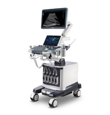
A transrectal ultrasound examination (TRUS) was performed on the Resona 7 ultrasound machine (Mindray, China). The volume of the prostate gland measured 36 ml, and a hypoechoic lesion of indistinct, uneven contour with a maximum diameter of 15 mm was seen in the peripheral zone of the right lobe, near the interlobular furrow (Fig. 1).

In power Doppler mode, an asymmetric increase in vascularity was seen within the hypoechoic lesion noted on B-mode TRUS imaging (Fig. 2).

Strain Elastography Findings
Using Natural Touch Elastography on the Resona 7 the stiffness of the prostate gland was assessed. The hypoechoic lesion within the peripheral zone did not show as significantly stiffer than the surrounding tissue with a Strain Ratio measuring between 0.56-0.87.

Contrast Enhanced Ultrasound Findings
For contrast enhanced ultrasound (CEUS), the area of interest was visualized immediately after injection of 2.4 ml of Sonovue (Bracco Swiss, SA, Switzerland) for 3 minutes in contrast mode. The hypoechoic lesion in the peripheral zone was shown to have hyperintensive contrast enhancement of relatively unchanged parenchyma, with rapid influx and outflow of contrast medium (Fig. 4).

Contrast Imaging Quantification Analysis
The time–intensity curve of the focal lesion was characterized by a rapid rise and subsequent rapid fall. The main quantitative indicators were TTP 16.5 sec., PI 35.7 dB, DT/2 78.6 sec., AUC 4634.2 dB/sec. For the intact peripheral zone, the time–intensity curve was characterized by a smooth rise and subsequent plateau phase, TTP was 63.9 seconds, PI 27.5 dB, AUC 4429.2 dB/sec. The DT/2 parameter was not determined (Fig. 5).

Biopsy Result
A systematic transrectal biopsy of the prostate gland was performed under ultrasound guidance with targeted sampling of 12 points.
Histological examination: in a column of tissue from the area of interest, the growth of acinar adenocarcinoma is 7 points according to Gleason (3 + 4). Histological result of focal formation of the peripheral zone on the right, acinar adenocarcinoma is represented by light tumor acinuses that do not have a basal cell layer. (Fig. 6).
Diagnosis
Malignant neoplasm of the prostate gland, T2сsN0M0.

Discussion
Classical ultrasound signs of prostate cancer are considered to be hypoechoic, hypervascular formation in the peripheral zone with a fuzzy contour [1]. These signs are characterized by low specificity, since similar changes can occur with various benign changes [2]. In this regard, multiparametric ultrasound is necessary to provide additional diagnostic information. Strain elastography can provide a map of the tissue stiffness in a given region of interest. While malignant lesions tend to be stiffer than normal tissue, this is not always so, as seen in this particular case where strain ratios did not indicate significant change in stiffness between the focal lesion and the surrounding intact tissue. CEUS can provide more detail regarding tissue vascularity. In this case, CEUS examination confirmed pathological vascularization within the focal lesion seen on B mode imaging, not just by qualitative signs, but also by quantitative parameters.
Reference:
[1] Ellis, W. J., Chetner, M. P., Preston, S. D. & Brawer, M. K., 1994. Diagnosis of Prostatic Carcinoma: The Yield of Serum Prostate Specific Antigen, Digital Rectal Examination and Transrectal Ultrasonography. The Journal of Urology, 152(5, part 1), pp. 1520-1525.
[2] Postera, A., Mischi, M., Rosette, J. d. l. & Wijkstra, H., 2015. Multiparametric ultrasound in the detection of prostate cancer: a systematic review. World Journal of Urology, 33(11), pp. 1651-1659.