Clinical information
A 56-year-old female patient was admitted to the hospital due to dizziness, asthenia, and pyrexia.
CBC results from Mindray BC-6000 Series
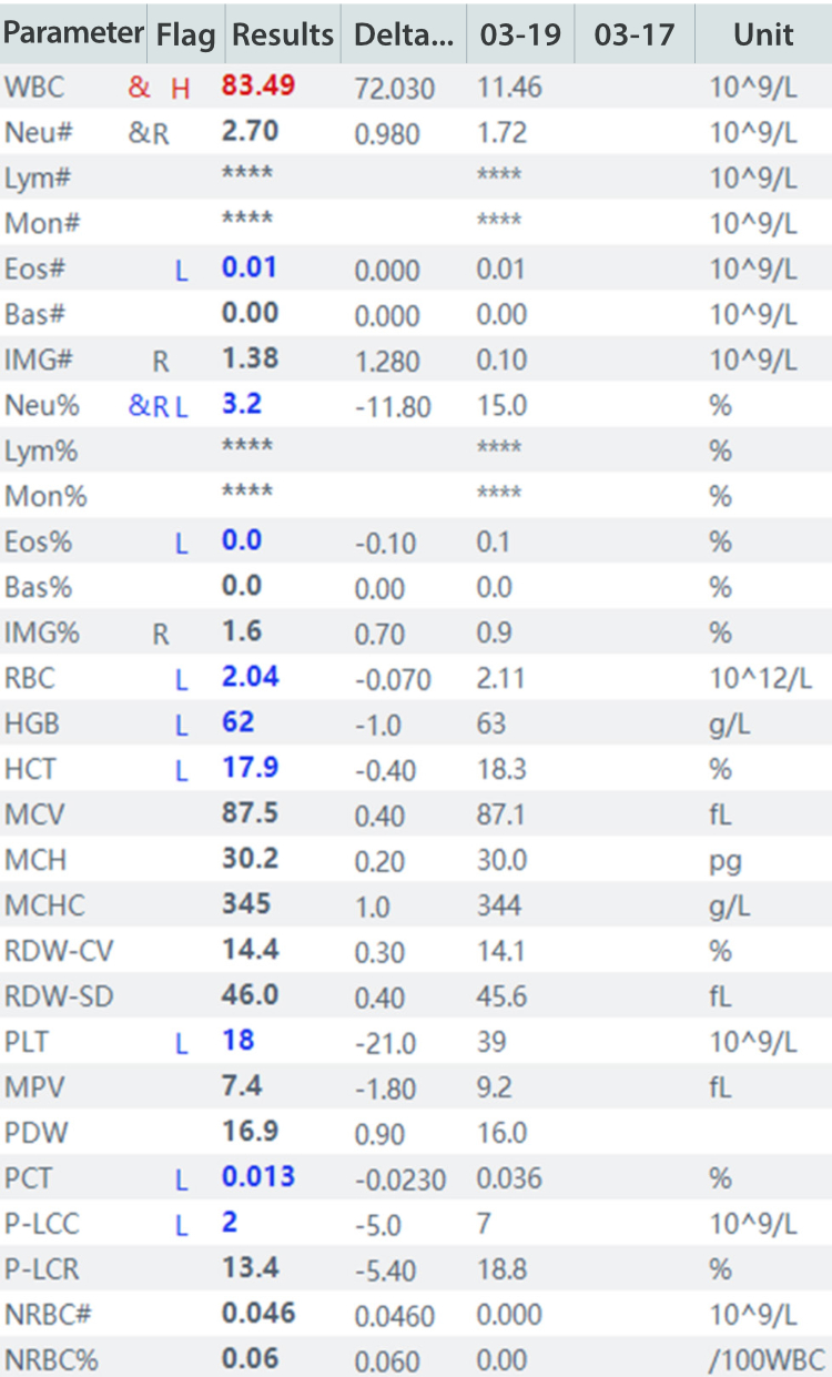
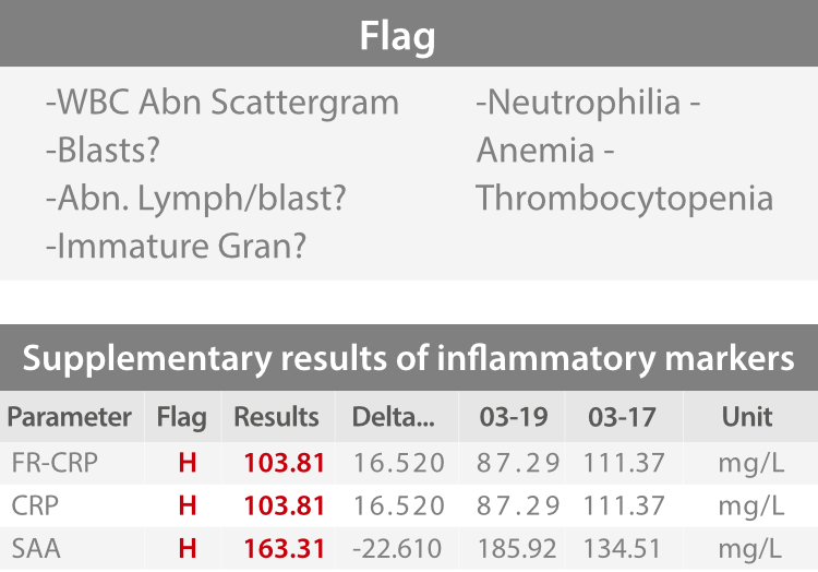
- High WBC and RBC indicating moderate anemia; low PLT was observed.
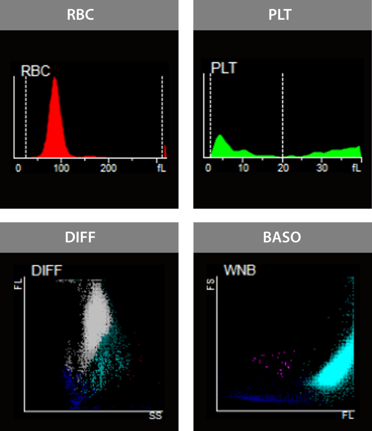
- The DIFF scattergram was abnormal with unclear and indistinguishable boundaries of cell groups; the overall scattergram was torch-like; the Monocyte area was dense and shifted up significantly; Neutrophil diffused upwards, with unclear boundaries from the Monocyte group. In the WNB channel, WBC shifted to the right, indicating the presence of a large number of promyelocytes.
- The re-examination rules were triggered, so the slides were prepared and being reviewed.
Peripheral blood morphology examination

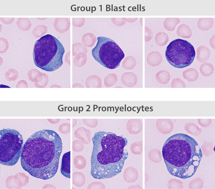
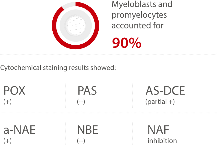
Results from manual re-classification: Blast cells and promyelocytes accounted for 95.5%, roughly divided into 2 groups according to cell morphology:
- Group 1 were round with large size. Their nucleus was large, light purplish-red, round or oval shaped, and located in the center or slightly offset to the center. The chromatins were fine and evenly arranged, flat as a thin layer of sand. The nucleoli were light blue and easily identified. Little cytoplasm was observed, presented in transparent blue or dark blue, with few or no particles.
- Group 2 was large cells in an oval or irregular shape. Their cytoplasm was abundant, presented in light gray to grayish blue, containing vacuoles and dust-like fine particles. The nuclei were round or oval. The chromatins were loose, but slightly coarser than those in the first group. The nucleoli were large and pronounced.
Bone marrow cytology test
About Acute Myelomonocytic Leukemia (AML-M4)
- Acute myelomonocytic leukemia (AMML) is a form of acute myeloid leukemia resulting from the proliferation of myeloblasts and mono-blasts, and accounts for 20% of AML.
- Some patients may experience fatigue, abnormal bleeding, anemia, and thrombocytopenia.
- Criteria for AMML diagnosis is more than 20% myeloblasts and promonocytes in the bone marrow.
- From marrow differentiation, proliferation of erythrocytes and megakaryocytes are inhibited.
Case analysis
M4 is a type of acute granulocytic leukemia and is divided into four subtypes, i.e., M4a, M4b, M4c, and M4Eo.

Invitation of Clinical Case Sharing
- Do you want to share your case with us? We will share your case in the upcoming issues with acknowledgement.
- IVD Cases: You are recommended to submit any typical, misdiagnosed, special or rare cases that received definite diagnosis or successful treatment. Cases that involved the communication between laboratory and clinical department are especially recommended.
- Format: You are required to include four sections in your entry: "Case Introduction, Case Details, Case Analysis, and an Executive Summary". The Case Details section must include: main medical history, positive test results, negative test results that are helpful for diagnosis, and diagnosis and treatment process.
- Please submit your entry in a Word document (in English). You need to send your entry by email to hoishan.kwok@mindray.com by April 30, 2023.
- The email should come in a subject that reads: country+name of the entry + name of the applicant.