


A special thanks for sharing this case to Dr. Nguyen Minh Thien of The Urology Department of Ho Chi Minh Medic Center.
Case Introduction:
A 71 YO male patient coming for prostate cancer screening, PSA levels higher than 100ng/ml. DRE showed an enlarged prostate with a hard nodule on the right lobe.
2D and Color Ultrasound findings:
Due to its anatomical location, it is recommended that a prostate ultrasound examination be conducted using real-time transrectal ultrasound (TRUS) with the highest frequency biplane endocavitary transducer (Mindray Resona i9, ELC13-4)
Prostate scanning was performed with a Mindray dedicated biplane convex-linear ultrasound probe. The transducer contains 2 ultrasound probes: the convex array and the linear array. The transverse image of the prostate was obtained with the convex array and the longitudinal image of the prostate was obtained with the linear array. Convex and linear arrays were supported with both Elastography techniques including: Strain Elastography (NTE, Natural Touch Elastography) and 2D Shearwave Elastography (SWE, Sound Touch Elastography).
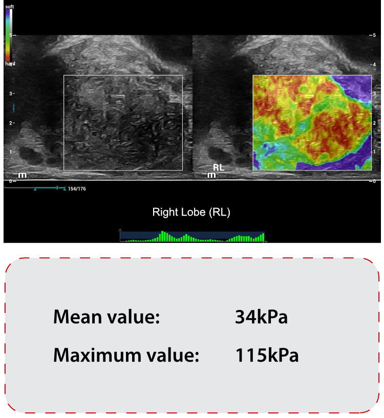
Ultrasound Findings of rigth lobe of the prostate:

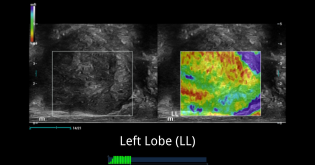
Ultrasound Findings of left lobe of the prostate:

Elastography Findings:
NTE – Strain Elastography:

SWE – Shear Wave Elastography:

The stiffness measurement results on PZ of RL were quite soft when compared with RL.
Biopsy Result:

A 6-core biopsy was done. Tissue sample numbers 1 to 3 were taken from the right lobe, and numbers 4-6 were taken from the left lobe.
The Biopsy Results Were
1,2,3: PROSTATIC ADENOCARCINOMA, Gleason score 7 (4+3), ISUP Gleason group 3 (C60): predominantly poorly formed/fused/cribriform glands with a lesser component of well-formed glands.
4,5,6: Normal prostate tissue.
Discussion:
Case introduction
PCa Screening on an elderly patient who had abnormal high PSA value (>100ng/ml) and abnormal DRE results (enlarged prostate and stiff nodule on RL)
Ultrasound
2D and Color Ultrasound showed a suspicious hypoechoic area in RL with hypervascularity and capsule-irregularity, but the LL findings were normal with just a bit uneven capsule, which made us confused whether the PCa happened on the whole prostate in both sides RL and LL or it just happen on the RL as RL has some typical findings of PCa.
Elastography
- NTE showed a very stiff area which was linked with hypoechoic area in 2D of RL. LL showed typically normal findings with a soft elastogram map and a well-defined border between TZ and PZ.
- SWE showed abnormal stiffness measurements of RL, which mean stiffness value was 34kPa and max stiffness value was 115kPa.
- While SWE showed normal stiffness measurements of LL, which mean stiffness value was 19kPa and max stiffness value was 36kPa.
Combined NTE and SWE results, we had more clinical evidence to confidently confirm that this patient had local PCa on RL, and LL of prostate seems to be normal.
Keynotes
- Elastography tool has an accurate and consistent result of stiffness difference between LL and RL, high correlation with PCa diagnostic.
- Combining NTE and SWE could help improve the characterization of prostate lesions and increase the detection of PCA.