Modern ultrasound techniques provide ever deeper insights into the human body with ever more detail. “The quality of sonography images is approaching the quality of MR images,” says Professor Dr. med. Christian Stroszczynski, Director of the Department for X-Ray Diagnostics at University Hospital Regensburg and adds that “the potential of sonography is far from being fully explored.” This is one reason why this study aims to expand the range of applications beyond the current use cases: in a multi-center project a new sonography technique using contrast agents for the diagnosis of liver tumors is being evaluated.
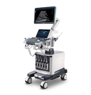
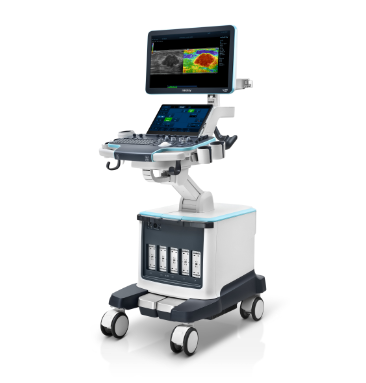
The ultrasound system being used in the study was perfomed on the Resona system updated to the latest R9 sofware. “The system others brilliant image resolution and minute detail within fractions of a second,” underlines Professor Dr. med. Ernst Michael Jung, Head of the Interdisciplinary Sonography Center at University Hospital Regensburg, and adds that “this allows us to get a quick and undistorted overview of the entire liver.” Moreover, the new software provides a penetration of up to 20 centimeters which is about twice as deep as with previous systems. A larger field of view facilitates orientation, recognition and spatial assessment. “Thus, we are now able to detect and reliably characterize small hepatocellular carcinoma,” Professor Jung summarizes his experiences so far.
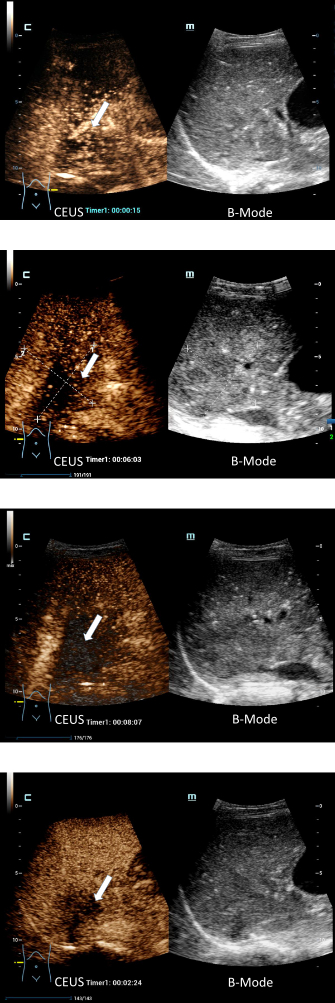
The use of contrast agents opens entirely new possibilities. Ten seconds after injection the contrast agent reaches the lesions which then brightly ‘glow’ for 30 seconds. In this time frame, the sonographer can get a good overview of the liver. Then the glow fades and after 3 minutes the lesions once again have the same color as the surrounding healthy tissue. This used to mark the end of the liver sonography exam with contrast agents.
The new Mindray software, however, does something striking: for four minutes the healthy liver tissue remains brightly colored while the tumor tissue gets darker and darker – thus the tissues once again can be clearly differentiated. “Previously, we were only able to examine the liver for three minutes”, which is a rather short time frame, “but now we get a second phase in which the tissue is shown in a light-dark effect. This strongly increases the quality of the clinical information and thus diagnostic confidence.” A specific post-processing feature allows the sonographer to brighten the malignant lesions in the first 15 seconds even more – and even darker in the later stages of the phase depending on the user preferences. Certain details can even be visualized in 3D post exam.
The exam can be documented in high-resolution video sequences and important still images can be highlighted – a feature that is a boon for all clinical disciplines. “To date, only single images are shown in tumor board sessions – and they can be difficult to assess for non-radiologists,” Professor Jung reports and adds that “the films of up to seven minutes are a major support tool for surgery planning. Even more: during surgery they can be used as a reference.”

These video sequences also play an important role prior to embolization where small tumors are cut off from the blood flow. “The images have almost the same quality as those that we acquire intra-surgery with the catheter. That means prior to surgery we see precisely which vessels the tumor uses,” Professor Jung explains. In the future, other organs such as pancreas or kidneys could benefit from this innovation. After all, Professor Jung concludes, “the objective of the current study is to expand the range of applications for this ultrasound technique.”
Today, however, the technique still has one major drawback: it requires high-performance ultrasound systems which are currently not available as mobile devices. Nevertheless, Professor Stroszczynski highlights another advantage: intuitive operation: “One reason why so far the potential of sonography has not been fully used is the non-intuitive operation of the devices. But recently handling the systems has become so simple that even less experienced physicians can quickly learn how to use them.”
Profiles
In October 2010, Professor Dr Christian Stroszczynski was appointed Chair of Radiology and Director of the Department for X-Ray Diagnostics at University Hospital Regensburg (UKR), Germany. Before, he had has been Deputy Director and Managing Consultant at the Institute for Radiological Diagnostics at Carl Gustav Carus University Dresden. From May 2016 to 2018 he chaired the German Society for Interventional Radiology and Minimally Invasive Therapy (DeGIR). In addition he served as Chair of the Working Group Ultrasound of the German Roentgen Society from 2014 to 2016 and was instrumental in developing the research focus ultrasound intervention. His work focuses on diagnostic and image-guided diagnosis and therapies for liver diseases, cancer and vascular medicine.
Professor Dr Ernst Michael Jung is Senior Physician and Head of the Ultrasound Center at UKR. Following this medial training at Saarland University and his specialist physician training at University Hospital Passau, in 2007 he was appointed Senior Physician at the Institute of X-Ray Diagnostics at University Hospital Regensburg. Jung is Professor for Translational and Experimental Sonography and active in national and international research projects on modern sonography techniques, particularly to improve tumor diagnostics and treatment. He is also member of different organizations such as DEGUM, AGUS and KVB as well as peer reviewer for all leading professional ultrasound journals.