Introduction
The patient, a 40-year-old woman, complained of a predominantly unilateral headache (debut from the forehead and periorbital area or occipital area), of a monotonous nature, which is accompanied by nausea, phonophobia, photophobia, osmophobia, increases with head turns. Headache has been bothering since the age of 16 with an increase in the frequency of seizures for the last 10 years. The triggers are the menstrual cycle and alcohol intake. When assessing the somatic and neurological status, pathology was not revealed. With MRI of the brain, there is no focal pathology. With ultrasound of brachiocephalic arteries (the study was conducted in a polyclinic) ‒ dissection of the left internal carotid artery or atherosclerotic plaque with 30% stenosis? The clinical diagnosis of a neurologist is "Migraine without aura".
Ultrasound examination - B- mode
Ultrasound examination (ultrasound) of the carotid and vertebral arteries was performed on the Resona 7 ultrasound machine (Mindray, China) using the L9-3U linear probe. When examined in B-mode in the area of bifurcation of the common carotid artery, a hyperechoic linear structure (slightly expanding at the base) with clear even contours, without signs of flotation, having a slope towards the internal carotid artery and protruding into the lumen of the vessel was visualized on both sides along the posterior wall: on the right by 1.5 mm, on the left by 3.1 mm (fig. 1). No structural changes were detected in other carotid and vertebral arteries.

In the bifurcation of the common carotid artery, markers indicate a linear hyperechoic signal with dimensions on the right (a) and left (b) in the longitudinal scanning plane. The type of hyperechoic signal (indicated by the arrow) in the left common carotid artery in the transverse scanning plane (c).
Ultrasound examination – Doppler modes
In the mode of color Doppler mapping, in the bifurcation of the left common carotid artery along the posterior wall, an anechoic zone with a smooth surface, about 1 cm long, was visualized (Fig. 2a), which was partially filled with blue blood flow (as opposed to red in the rest of the carotid arteries) (Fig. 2b). The "aliasing" effect over the zone of detected changes was not detected. In the spectral Doppler mode, in the bifurcation of the common carotid artery, the velocity parameters of blood flow were within normal limits, no areas with local changes in hemodynamics were detected (Fig. 3).
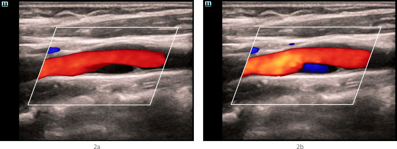
In the bifurcation of the common carotid artery along the posterior wall, an anechoic structure is visualized, having a hyperechoic contour in the proximal shoulder, and partially filled with blue blood flow during different phases of the cardiac cycle and when the device settings are changed.
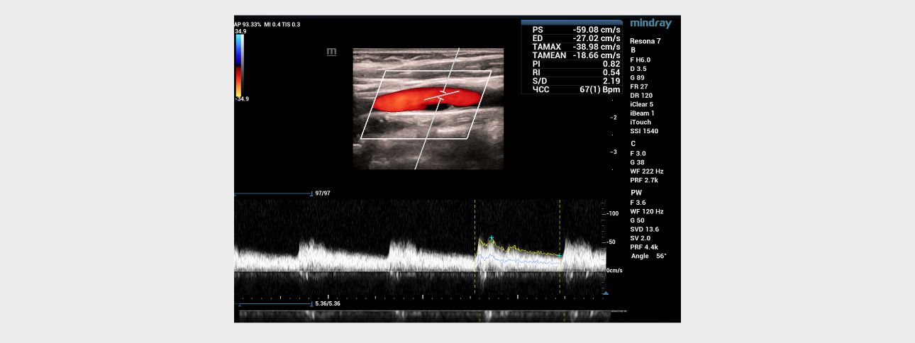
Blood flow with normal velocity parameters (Vs=59 cm/s) is recorded above the anechoic area.
Ultrasound examination – vector analysis of blood flow mode (V Flow)
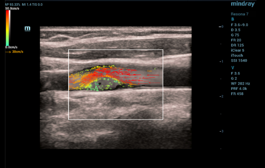
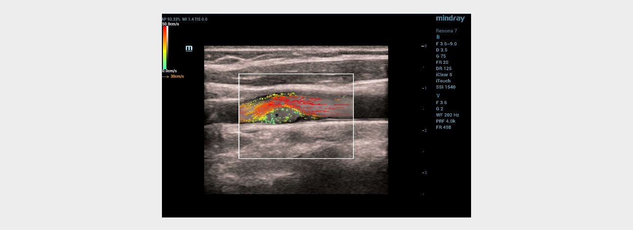
When the vector analysis mode (V Flow) was activated, the complete filling of the lumen of the left common carotid artery in the bifurcation area with colored vector arrows was clearly traced. The following changes were registered immediately after the linear hyperechoic structure: 1) shorter color arrows compared to arrows from the main blood flow in the vessel; 2) the arrows had a different color pattern (green, blue, yellow) in contrast to the red and orange arrows of the main stream; 3) the short arrows had a multidirectional, vortex-like direction in contrast to the laminar flow, which was determined over the pathological area of the vessel with a hyperechoic linear structure (Fig. 4). Given the absence of signs of flotation of hyperechoic linear formations and the topical symmetry of the location, it was assumed that these changes are characteristic of the carotid web.
Computed tomography angiography
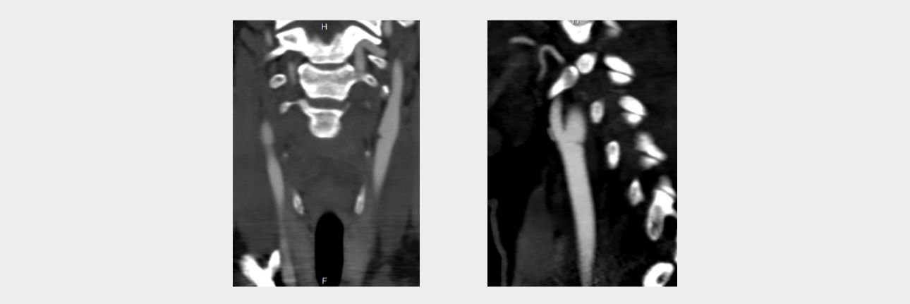
In order to clarify the changes detected on ultrasound, the patient underwent computed tomography (CT) angiography of extracranial arteries with intravenous contrast with Omnipack 350 in an amount of 50 ml on the SOMATOM Definition AS device (Siemens, Germany). According to the results of the study, linear structures were determined in the bifurcation of the common carotid artery on both sides, leading to a shelf-like defect in filling the vascular lumen in the sagittal scanning plane, more pronounced on the left (Fig. 5).
Conclusion: CT angiographic signs of the minimal variant of fibromuscular dysplasia (Carotid Web).
Discussion
Initial examination
The absence of specific complaints and focal neurological symptoms associated with a stenosing lesion of the carotid sinus on both sides did not allow clinically suspecting pathology in the carotid arteries at the extracranial level. Complaints, anamnesis of the disease and belonging to the female sex gave grounds for the diagnosis of "Migraines without aura (simple migraine)". However, in order to assess the condition of the blood vessels supplying the brain, the patient was assigned an ultrasound of the brachiocephalic arteries, in which dissection of the left internal carotid artery was suspected. To clarify the nature of the detected changes, the patient was sent to a specialized institution dealing with vascular pathology of the brain (Federal State Budgetary Scientific Institution "Scientific Center of Neurology").
B-mode, Dopplerography
The results of ultrasound in the FSBI NCN. According to seroscale and Doppler ultrasound modes, hyperechoic linear structures were detected in the bifurcation of the common carotid arteries on both sides, with a large narrowing of the vessel lumen on the left, which could be a sign of intimal detachment and suggest dissection or a local form of fibromuscular dysplasia. An anechoic staining defect of the vessel lumen was detected behind the hyperechoic structure in the distal direction on the left in the CDK mode, which was partially filled with the color (blue) opposite to the main flow direction when changing the settings of the device and the phase of the cardiac cycle. Since it was not possible to completely paint the anechoic defect with a color stream, while not flooding the lumen of the common carotid artery with adjacent structures with color, it was impossible to exclude the presence of an atherosclerotic plaque in a typical place of its formation with a tire visualized in its proximal part or, but less likely, a parietal thrombus. The absence of an increase in the rate of blood flow and local changes in hemodynamics indicated the hemodynamic insignificance of structural changes in the carotid arteries on both sides.
Ultrasound vector analysis of blood flow
The V Flow mode made it possible to clearly see the full filling of the vessel lumen with colored arrows directly behind the hyperechoic linear formation on the left. The direction, color and size of the arrows in this area of interest clearly provided information about the slow-speed, retrograde and then vortex-like flow of blood in the vessel, which explained the poor color filling of the vessel lumen directly behind the hyperechoic linear structure in the color Doppler mapping mode, due to the high dependence of the latter on the speed and direction of moving blood particles (erythrocytes). The technology of vector analysis of blood flow made it possible to exclude the presence of an atherosclerotic plaque or a parietal thrombus.
Thus, ultrasound differential diagnosis was carried out between dissection with detachment of intima and a local atypical form of fibromuscular dysplasia by the type of carotid web. The symmetry and non-typical location for spontaneous dissection of hyperechoic linear structures protruding into the lumen of the vessel, the absence of indications of traumatic neck injury (surgery, fall, etc.), and, most importantly, the absence of signs of their flotation, indicated the rigidity of the formations and excluded the stratification of the vascular wall.
Computed tomography angiography
The results of the study confirmed the conclusion of ultrasound that the patient has a carotid network on both sides with a large stenosis on the left.
Key points
The carotid web is a rare form of focal fibromuscular dysplasia, which is a thin protrusion of the intima of a linear shape, most often localized in the sinus region of the internal carotid artery [1,2]. The carotid web protrudes into the lumen of the carotid artery, causing local disruption of blood flow and the formation of blood clots, which can lead to subsequent ischemic stroke [3]. Currently, CT angiography is the preferred method of noninvasive imaging for the diagnosis of the carotid web, but ultrasound in combination with CT angiography or magnetic resonance imaging complement each other in the characteristics of carotid webs [4]. Wide awareness and alertness of ultrasound diagnostics specialists regarding this pathology can contribute to early diagnosis and prevention of fatal cerebral complications.
In the presented patient with migraine headaches, ultrasound revealed asymptomatic structural changes of the same type (more pronounced on the left) in the area of bifurcation of the common carotid arteries, leading to hemodynamically insignificant stenosis of the vascular lumen. A comprehensive ultrasound assessment, including modern V Flow technology, which provided very important information about the local characteristics of blood flow in the vessels, made it possible to establish a diagnosis of the "Carotid web", which was subsequently confirmed by CT angiography data. Taking into account the hemodynamic insignificance, this gave reason to adjust the treatment by adding an antiplatelet drug (clopidogrel 75 mg once a day, for a long time) to the main antimigrenous therapy for the prevention of thrombosis in the carotid web area and take the patient under regular (annual) dynamic ultrasound observation.
Reference
1. Gouveia EE, Mathkour M, Bennett G, et al. Carotid Web Stenting // Ochsner J 2019; 19(1): 63-66. doi: 10.31486/toj.18.0143.
2. El Mesnaoui R, Nikiema S, Massimbo D, et al. The carotid diaphragm, an often overlooked cause of stroke by cardiologists // J Surg Case Rep 2022; 2022(7): rjac350. doi: 10.1093/jscr/rjac350.
3. Guglielmi V, Compagne KCJ, Sarrami AH et al. Assessment of Recurrent Stroke Risk in Patients With a Carotid Web // JAMA Neurol. 2021. Jul 1; 78(7): 826-833. doi: 10.1001/jamaneurol.2021.1101.
4. Zhu С, Li Z, Ju Y et al. Detection of Carotid Webs by CT Angiography, High-Resolution MRI, and Ultrasound // J Neuroimaging. 2021. 31(1): 71-75. doi:10.1111/jon.12784.
