Many laboratories are troubled by problems such as lengthy analysis processes and less efficient microscopic examinations due to lacking of experts in cell morphology and inadequate equipment. This makes them hard to reach a 30% overall re-exam rate, a target recommended by the international consensus group for hematology review. Furthermore, the abilities of the laboratory staff are varied, leading to inconsistency in lab standards and results.
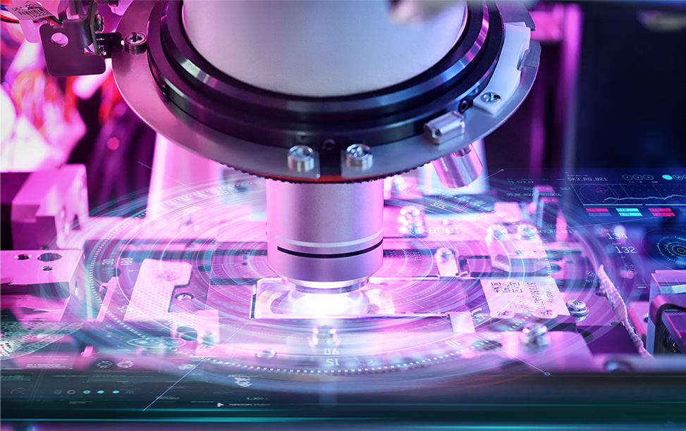
Mindray’s MC-80 Automated Cell Morphology Analyzer is designed to provide "More Clarity, More Intelligence, and More Productivity" for cell morphology analysis, with intelligent tools to help discover the truth about cells.
Committed to improving the efficiency and accuracy of laboratories, Mindray has been collaborating with experts and scholars in hospitals around the world since 2014. Based on intensive research, Mindray has come up with the innovative solutions in cell imaging such as Multi-layer fusion technology, "Solid Rock" Hyper-stable Anti-shake System, Fly-mode Technology, and many more.
More Clarity: more details beyond appearances
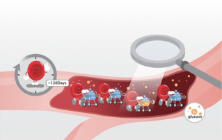
Cells are the basic units of life, and cell morphology test is one of the prime tools for detecting malignant diseases. Pathologists study the internal structure and details of the cells for various needs.
Currently in most laboratories, morphology experts observe the cell images of each re-examination sample manually with a microscope, making it a prolonged process when the test volume is large.
In order to effectively alleviate the workload of manual analysis with a microscope, cell morphology analyzers need to simulate the details captured by manual microscopy as clearly as possible. How can planar imaging of cells achieve this?
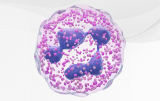
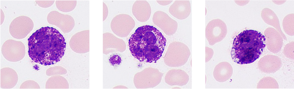
The MC-80 innovatively captures 20 images of each cell at different depths of field.
The essence of the multi-depth fusion technology is to recreate manual focusing normally performed by the doctor by superimposing the clear parts of the 20 images. In the cell imaging from the MC-80, the features and internal details of normal and abnormal cells can be clearly presented.
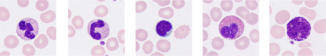
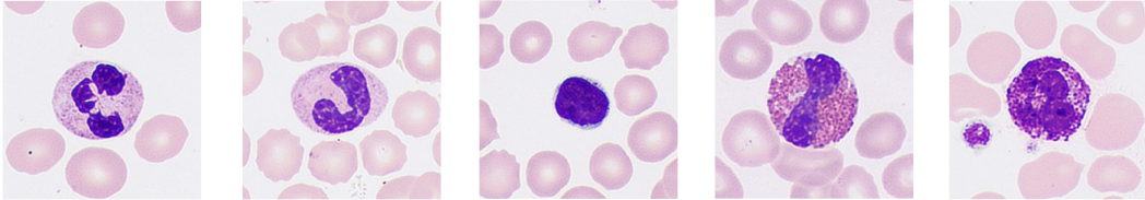
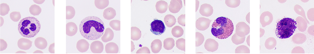

Therefore, doctors can gain insight into the pathological features of cells for more accurate early screening of blood disorders and infectious diseases, avoiding misdetection.
The essence of the multi-depth fusion technology is to recreate manual focusing normally performed by the doctor by superimposing the clear parts of the 20 images. In the cell imaging from the MC-80, the features and internal details of normal and abnormal cells can be clearly presented.
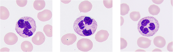
Segmented neutrophils
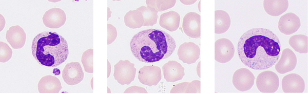
Band neutrophils
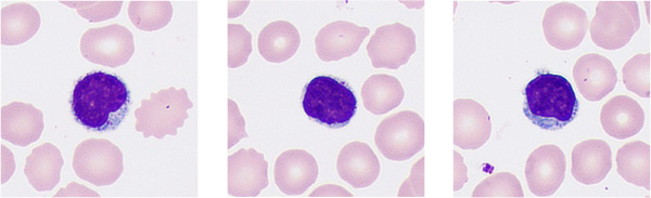
Lymphocytes
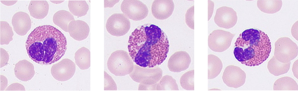
Eosinophils
Basophils
Therefore, doctors can gain insight into the pathological features of cells for more accurate early screening of blood disorders and infectious diseases, avoiding misdetection.
More Intelligence: delivering an all-round view of samples

Among the complex information, disordered information is invalid and creates a barrier between doctors and the truth.
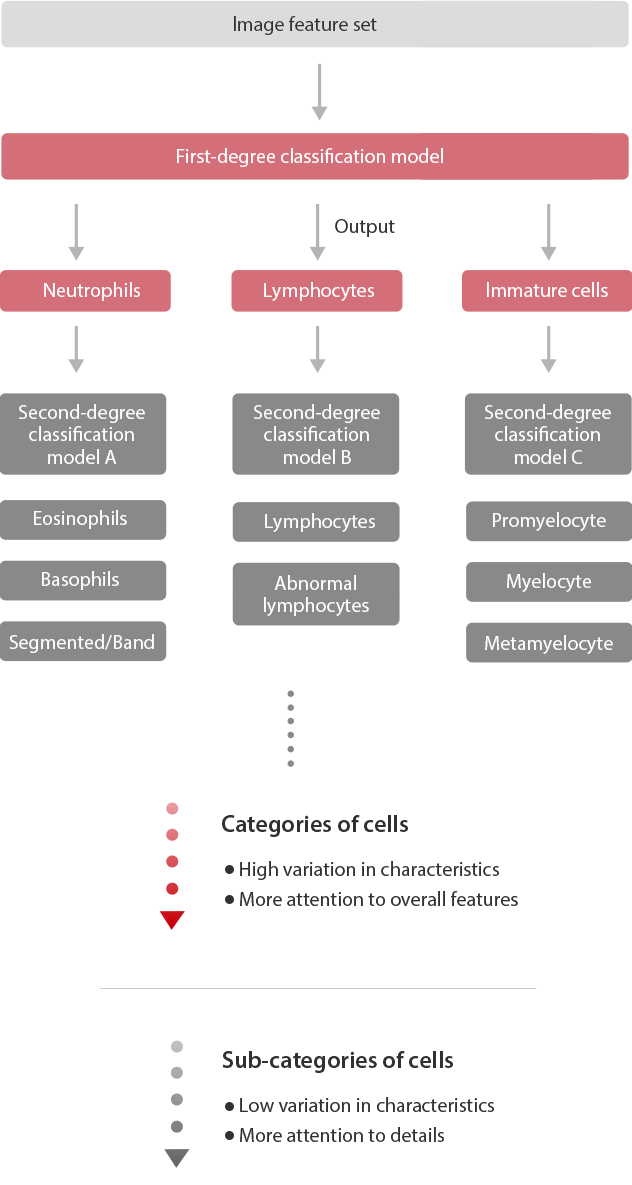
Collecting cell images is just the beginning. It is then necessary to classify each cell correctly to reflect the all-around view of the sample. When analyzing manually, the classification process is often recorded one at a time by the doctor. However, as the various types of cells are similar, doctors can only classify them based on their details. Over time, errors occur, affecting the accuracy and efficiency.
Through reliable identification and pre-classification of different types of cells, the MC-80 can accurately reflect the all-around view of the sample, thus helping doctors to understand the status of the patient.
Accurate recognition is based on the accurate extraction of features from the digital images of cells. With the guidance of morphology and pathology experts in the early stage, Mindray team extracts the information regarding cell color, texture, and geometric characteristics on different scales. By improving the recognition rate of cells using cascade classification, pathologists can accurately confirm the types of cells through comparison within categories and subcategories.
Improve cell recognition rate by cascade classification
The MC-80 can intelligently adjust the analysis mode before and during the process according to the information from the cellular analysis line to improve diagnostic efficiency.
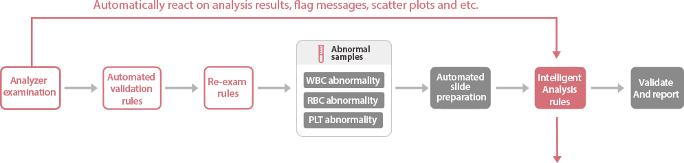
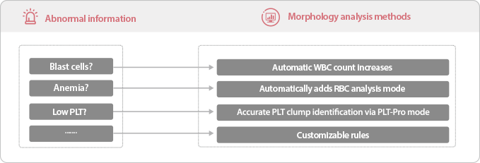
For the false decreased platelet counts due to clumping, the MC-80, with its unique high-speed FLY-MODE, can scan the body, both sides, and tail of the smear within one minute, accurately identifying platelet clumping, avoiding time-consuming manual confirmation.
More Productivity: improving the efficiency of morphology analysis
Until now, under stable lighting, capturing a clear image requires long exposure time and almost no camera movement. It seems that speed and clarity can’t be achieved at the same time.
The same goes for cell morphology analysis. In addition to clarity, efficiency is also important for finding the truth faster. Traditional manual analysis takes 5-8 minutes for each sample. In large laboratories, the huge workload requires specialized personnel, resulting in high labor costs.
Laboratory has to review large quantities of samples.


1.5+ hours 
Total analysis time for MC-80

Only by completing the test faster can the microscopic examination be further popularized. Increasing cell morphology testing efficiency is a major goal in the pursuit of truth.
The MC-80 consistently captures cell images at a high speed, processing one sample per minute. Through flexible single-slide sampling, it can quickly generate results for urgent cases.
Compared with traditional cell analyzers, the MC-80 has many breakthrough features.
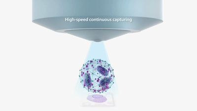
Total quantity of images taken per hour

120000
MC-80’s capturing performance
MC-80 is equipped with a built-in SOLID ROCK hyper-stable anti-shake system. After repeated verifications, the new aerospace material can provide the stiffness required by the MC-80 during high-speed motion. The stable structure and advanced high-frequency exposure algorithm can address the small horizontal and vertical perturbation to ensure fast, accurate, and reliable cell analysis.
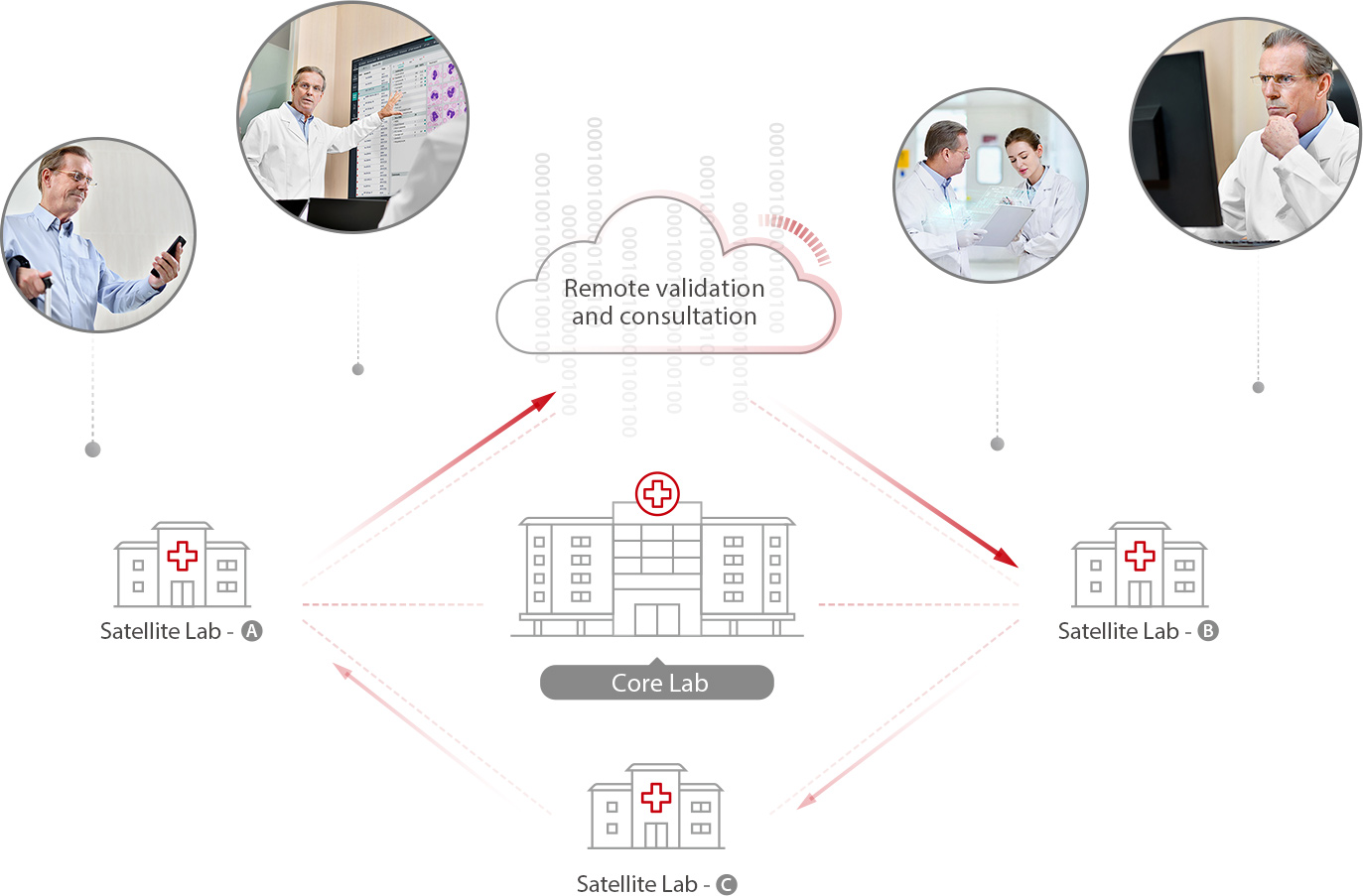
In case of technical problems during the examination, the MC-80 supports multi-terminal remote validation, allowing consultation to be immediately available.
Remote review and consultation in multiple locations
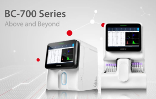
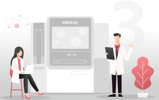
MC-80
Automated Digital Cell Morphology Analyzer
More Clarity, More Intelligence, More Productivity.
Mindray’s MC-80 Automated Cell Morphology Analyzer provides more accurate and reliable morphology analysis with its industry-leading technology in the field. By addressing the key challenges in morphology analysis, Mindray provides an comprehensive auto hematology analysis solution to empower trust, delivering accurate results while meeting laboratories’ demands on efficiency worldwide.