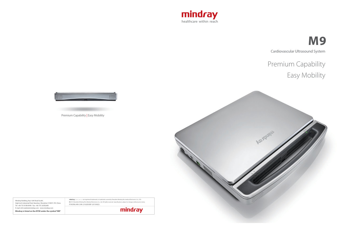
Based on Mindray's new generation ultrasound platform, mQuadro, M9 has raised the industry standards to an all new level. Advanced signal transmission and reception processors provide highly sensitive and accurate echo detection. Innovative transducer technologies allow for better penetration, higher resolution, greatly enhancing your diagnostic experience.

Echo-enriched Beam Forming
Echo-enriched beam former permits the use of traditionally neglected echo signals of adjacent beams to form one finer and stronger imaging beam, providing better ‘out-of-focus’ image resolution and deeper image penetration.

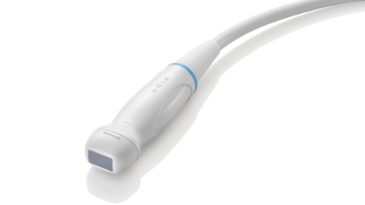
3T Transducer Technology™
Mindray’s patent transducer technology to increase image bandwidth and transmission efficiency.
· Triple-matching layer design for higher sensitivity, wider bandwidth, and improved S/N
· Total-cut design for lower cross-talk noise, better directivity, and improved lateral resolution
· Thermal-control design for better acoustic transmission

Multi-Beam Formation
Maximum 12 times tasking for one transmitted beam, resulting in excellent time resolution and higher frame rate.

iClear (Need application specific images)
Gain improved image quality based on auto structure detection.
· Sharper & Continuous Edges
· Smooth Uniform Tissues
· Cleaner ‘no echo areas’
iBeam™
Permits use of multiple scanned angles to form a single image, resulting in enhanced contrast resolution and improved visualization.

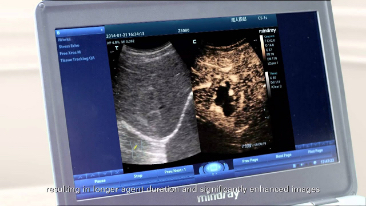
PSHI™ (Phase Shift Harmonic Imaging)
Purified Harmonic Imaging for better contrast resolution providing clearer images with excellent resolution and less noise.

FCI (Frequency Compounding Imaging)
Permits compounding of different frequencies to form best whole field image homogeneity, providing better penetration especially for high frequency scan.

Free Xros M™
Gain precise anatomical observation by freely placing sample lines at any angle. Attain better images through simultaneous display of up to 3 sample lines.

Free Xros CM™
Accurately evaluate myocardial motion at different phases, and simultaneously determine myocardial synchronization. High frame-rate providing you with accurate results:

TDI
Tissue Doppler Imaging allows you to quantitatively evaluate local myocardial movement and function, providing complete TDI modes for faster and direct diagnoses.

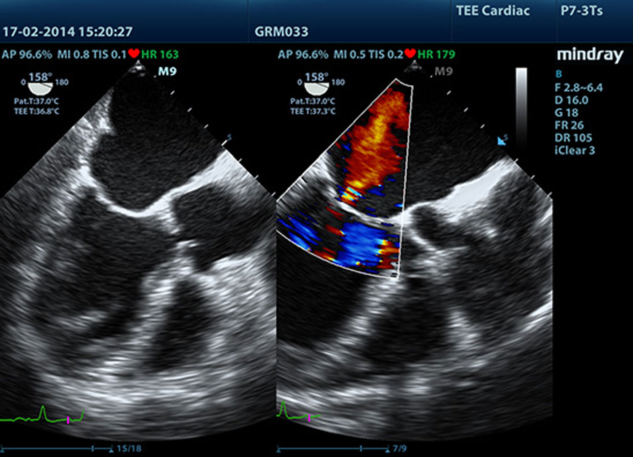
LVO with Stress Echocardiography
M9’s premium capabilities allow for LV opacification during stress, enhancing discrimination between myocardial tissue and blood pool, providing better visualization of the endocardial surface. Stress Echo feature on M9 includes a complete package for pharmacological stress and exercise stress echo. The package is supported by a flexible reporting system that can be optimized for your individualistic needs.
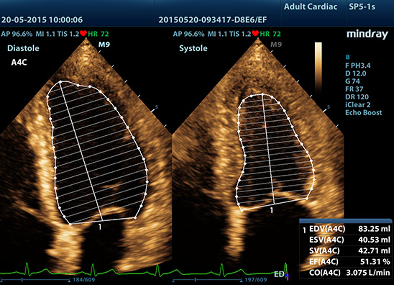
AutoEF
Intelligent way to analyze 2D echo clips to automatically recognize diastole/systole frames and output EDV/ESV/EF etc. results by Simpson method.
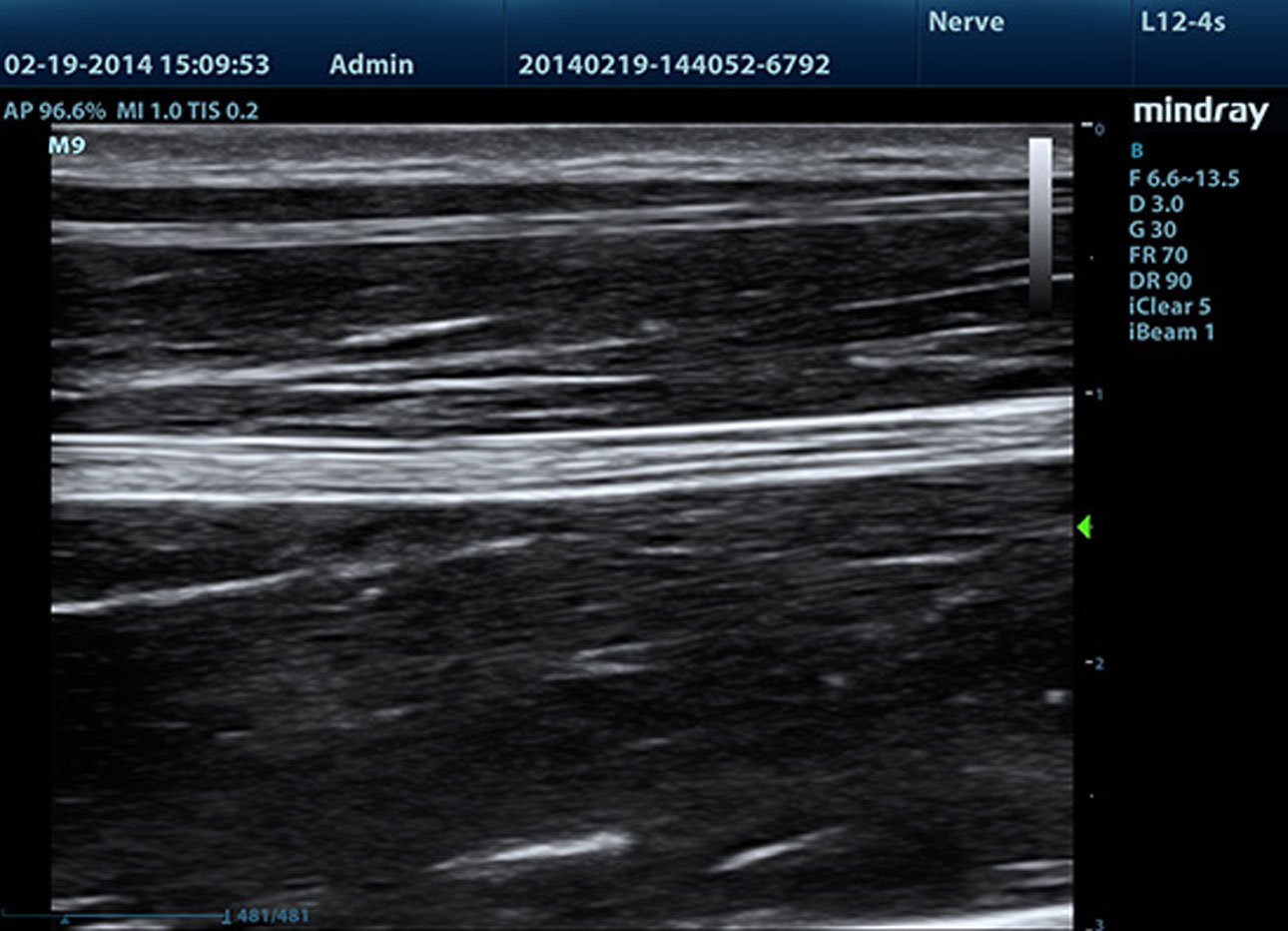
iNeedle™ (Needle Enhancement Visualization)
Advanced needle visualization allows the needle increased visibility even during steep-angled procedures, while maintaining superior image quality. Improved confirmation of needle location in tissue minimizes harm to surrounding tissue.
Raw Data
Enables optimum flexibility for post processing of the stored images including parameter adjustments, adding comments and measurements, allowing maximum productivity during scanning.

Auto IMT (Intima-Media Thickness)
Auto measurement of anterior and posterior wall thickness providing accurate carotid status.

Auto LV
Simple measurement procedure for left ventricle enhanced by auto-trace functionality and easy manual correction.

iStation™
Mindray’s unique Patient Information Management System allowing you to integrate, review, archive and retrieve patient data effectively.
iTouch™
Gain instant auto image optimization in B, Color and PW Modes on the click of a single key.

iWorks™
A smart tool to let you focus more on the patient.Helps in significantly reducing the patient scan time through standardization and user-defined capability.

iScanhelper
Dedicated inbuilt tutorial software, iScanHelper provides you with an interface to learn “How & What” of ultrasound scanning. Supported with excellent practical examples and illustrations, iScanHelper, with its easy and practical usability, is an effective educational tool either for beginners or experienced professionals.