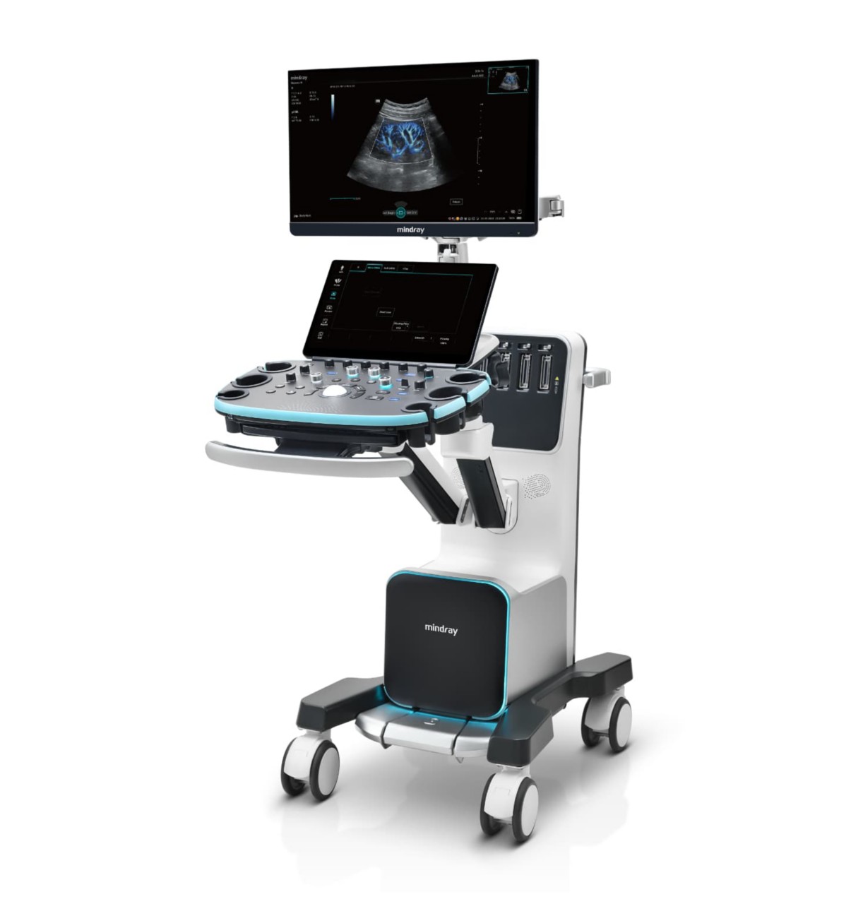
Premium Ultrasound System for Radiology

Resona A20

Reveal What Matters

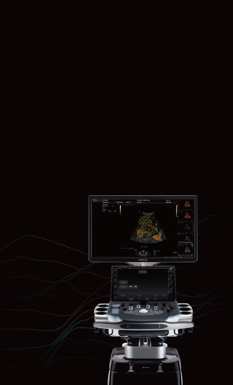
Resona A20 product image

Premium Ultrasound System for Radiology

Resona A20

Reveal What Matters

Resona A20 background image

Reveal What Matters

By revealing the unseen, we empower you to embark on a transformative journey of discovery and understanding, enabling a deeper comprehension of human body and the world around us.

Driven by this mission, Mindray is about to release the premium ultrasound system - Resona A20. Powered by the Acoustic Intelligence Technology platform, it has pushed ultrasound imaging performance to a new level, helping clinical experts to achieve accurate diagnosis and academic exploration. Together, we can explore new horizons and push the boundaries of medical knowledge.

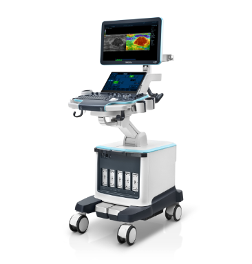
Resona A20 product image with background

AIT Platform

Acoustic Intelligence Technology

The AIT Platform has achieved significant advancements in acoustic and electrical data, imaging algorithms, and system computing power. High-quality acoustic and electrical data are ensured by the AFM Transducer and FreeWave Tx/Rx Technology. Additionally, FreeBeam Beamforming and FreeIQ Intelligent Image Processing technology are dedicated to faithfully revealing tissue details.

transparent AIT Platform icon
AFM Transducer
FreeWave Tx/Rx
Hyper Parallel Computing
FreeBeam Beamforming
Free IQ Image Processing
Data
Computing
Power
Algorithm

Precise Imaging Diagnosis

Based on the AIT platform, Resona A20 provides clinicians with superior ultrasound imaging clarity for difficult clinical disease diagnosis. HD Scope+ is based on the innovative adaptive beamforming technology, which further reveals the tiny details of lesions with powerful ultrasound diagnostic capabilities.

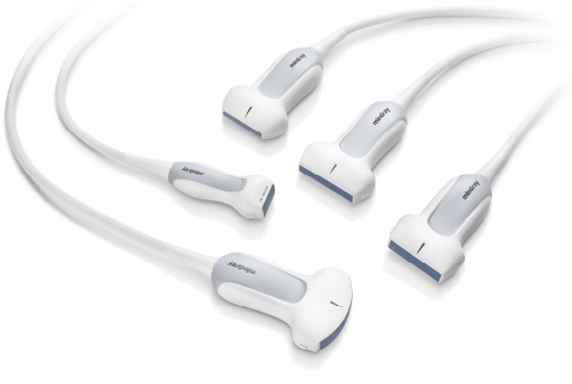
AFM Transducers

Mindray's next-generation transducers incorporate advanced technologies to enhance energy conversion efficiency, provide ultra-wideband coverage, and improve acoustic focusing capabilities, ensuring precise clinical diagnoses.

AFM Transducers
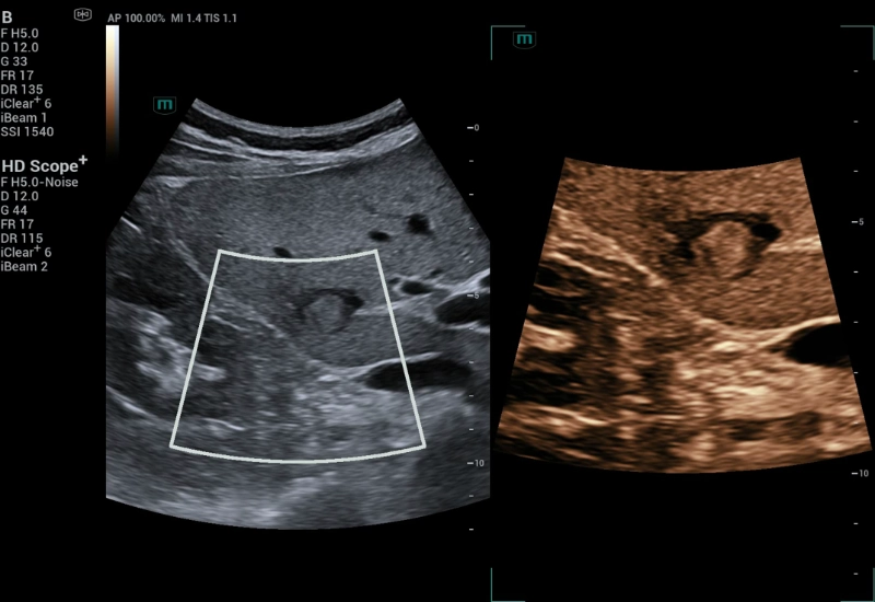
HD Scope+

Powered by FreeBeam beamforming technology from the AIT platform, HD Scope+ can extract more effective echo information. Depending on specific clinical needs, target-focused image enhancement is achieved using FreeIQ processing technology. HD Scope+ and B-mode provide dual live imaging, revealing intricate details of lesions for deeper clinical insights.

HCC, HD Scope+

HCC

Resona A20 scan for Thyroid Nodule

Thyroid Nodule

Resona A20 scan for Breast Lesion

Breast Lesion

UMA
HiFR CEUS
Sound Touch Elastography

The newly upgraded UMA captures minute low-speed blood flow with high sensitivity, high spatial resolution, and excellent motion artifact control. This enhancement has the potential to significantly improve diagnostic efficiency for organ perfusion evaluation and tumor research.

High sensitivity

High resolution

Better motion artifacts control

Transplanted Kidney | sUMA

Skin Wart | pUMA

HiFR CEUS offers ultra-fast imaging compared to traditional methods. By capturing detailed perfusion in the arterial phase, it enhances tumor diagnosis and the study of perfusion morphology.

6-8 times faster CEUS

Study on perfusion morphology of tumors

More clear perfusion details in the arterial phase

Metastatic Liver Carcinoma | HiFR CEUS

STE pushes the boundaries of image performance. With multiple quality control and intelligent tools, it intuitively and quantitatively evaluates tissue stiffness, making it highly effective for liver brosis and breast tumor assessments.

Superior imaging performance

Multiple quality control tools

Smart tools

Breast Carcinoma, STE

Breast Carcinoma | STE

M Reference

M Reference is a multi-parametric combined analysis tool that enables real-time, same-slice, and same-screen MPUS diagnosis. Unlike traditional single ultrasound imaging, it offers multi-dimensional diagnostic information and quantitative evaluation indicators for diseases.

Multi-parametric combined analysis

Multi-parametric quantification tools

Real-time, one-screen assessments

Breast Mass, M Reference

Breast Mass

Fatty Liver, M Reference

Fatty Liver

Multi-parametric quantification tools

Multi-parametric quantification tools

Innovative Clinical Research

Equipped with a wide range of innovative imaging technologies, the Resona A20 supports clinicians in cutting-edge clinical research. Super Resolution CEUS (SR CEUS) reveals blood perfusion details at the micron level, aiding clinicians in the exploration of early microcirculatory changes in lesions. Additionally, STVi shear wave viscoelastography, a novel technique for assessing tissue viscosity, demonstrates great potential for studies on chronic liver diseases and tumors.

Super Resolution CEUS
STVi
Quantitative Vascular Analysis Tools

Powered by the AIT platform, the Resona A20 delivers an all-in-one integrated solution for super-resolution imaging, a capability previously difficult to achieve. SR CEUS reveals the intricate microcirculation details of lesions at the micron level, aiding in microcirculatory perfusion studies in oncology.

Micron level resolution

Microvascular detection capabilities

Quantification tools

Focal Nodular Hyperplasia

Focal Nodular Hyperplasia | Density Map

Focal Nodular Hyperplasia Blood Flow Density Map

Focal Nodular Hyperplasia | Direction Map

Focal Nodular Hyperplasia

Focal Nodular Hyperplasia | Velocity Map

STVi enables the quantitative evaluation of tissue viscosity and provides real-time multi-parameter imaging, offering a more comprehensive approach to imaging diagnosis and quantitative analysis of chronic liver diseases, breast lesions, and other conditions.

Dual quantitative coefficients

Chronic liver disease assessment

Multiple quantification tools

Breast tumor assessment

Breast Lesion STVi

Breast Lesion

Fatty Liver STVi

Fatty Liver

The Resona A20 introduces a new generation of vascular quantitative analysis tools, featuring RF-data-based vascular pulse wave velocity and wall shear stress analysis. These advancements aid in the assessment of arterial vascular sclerosis.

Holo-PWV

V Flow and wall shear stress analysis

Carotid Artery Holo-PWV

Carotid Artery | Holo-PWV

Carotid Artery V Flow

Carotid Artery | V Flow

A New Level of Image Clarity

{{ item.title }}
{{item.title2}}

false