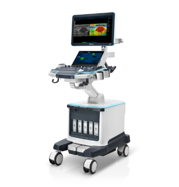
Recho R9
Clarify the Echo - Seeing Beyond the Beat


Clarify the Echo - Seeing Beyond the Beat
Facing the increasing population of cardiovascular patients, the Recho R9 series offer accurate solutions for diagnosing heart diseases by high-resolution cardiac structure imaging, precise cardiac function quantitative assessment and efficient workflow.

Innovative Echocardiography Platform - eZST+
Leveraging zone sonography technology, this innovation solves the conventional limitations of ultrasonic beam synthesis in spatial and temporal resolution. It achieves superior imaging velocity, higher frame rates, enhanced cardiac flow sensitivity, and exceptional uniformity across the acoustic field. Compatible with matrix transducers, the system enables real-time 3D cardiac imaging capabilities. This advancement facilitates meticulous observation and evaluation of cardiac anatomical structures.

Macro-array Beamforming Technology
Accurate transmitting and receiving of 3D acoustic field
Ultra-Dynamic Range Flow
Compatible display of abnormal high-speed flow and low-speed flow
Broaden Zone Harmonic Imaging
Synchronous improvement of image frame rate and detail resolution
Auto Touch
Real-time image optimization

Clear Imaging
Accurate Quantification
Real-time 3D Cardiac Imaging
Easier to observe the spatial shape and movement of cardiac structure

X-Vue
iLive Echo
Stress Echo
Under pharmacological, exercise, or other stress conditions, assess the coronary blood flow reserve capacity of patients to evaluate the extent of myocardial ischemia and myocardial viability

Intelligent Empowerment
Automated Cardiac Function Analysis

Auto Strain LV
Auto Strain LV intelligently identifies and tracks tissue movement for rapid, efficient myocardial motion evaluation

Auto EF
Auto EF automatically identifies the type of sections and providing an assessment of the left ventricle's overall systolic function
Intelligent Empowerment
Automated Cardiac Function Analysis

Auto VQLA
Auto VQLA automatically identifies the type of sections and tracks the left atrial wall, aiding in the computation of left atrial volume and volume index

Auto DFR
In spectral and tissue doppler modes, sampling volume is automatically positioned at the mitral valve orifice for spectral display and measurement, aiding in assessing left ventricular filling pressure and diastolic function
Intelligent Empowerment
Quantitative analysis of MCE
Post-flash analysis records contrast medium reperfusion rate and intensity

Echo Contrast Imaging QA
Intelligent Empowerment
Vascular Function Assessment

V Flow
A novel approach for vascular hemodynamic analysis, using color coded vector arrows to display flow velocity magnitude and direction

RIMT
RF-Data based Intima-Media Thickness, provide precise IMT thickness measurement in real time

R-VQS
RF-data Based Quantitative Analysis on Vessel Stiffness, provide an precise and quantitative tool for vascular stiffness evaluation
Clear Imaging
Movement of Cardiac Structures
Delivering high frame rates, exceptional detail resolution and high flow sensitivity

Rheumatic heart disease

Atrial septal defect
Real-time 3D Cardiac Imaging
Under pharmacological, exercise, or other stress conditions, assess the coronary blood flow reserve capacity of patients to evaluate the extent of myocardial ischemia and myocardial viability

X-Vue

iLive Echo
Contrast-enhanced Echocardiography
Enhances left ventricular imaging clarity, enables precise evaluation of left ventricular function and myocardial perfusion

LVO

VLMI Contrast
Stress Echo
Under pharmacological, exercise, or other stress conditions, assess the coronary blood flow reserve capacity of patients to evaluate the extent of myocardial ischemia and myocardial viability

Accurate Quantification
Intelligent Empowerment
Automated Cardiac Function Analysis

Auto Strain LV
Auto Strain LV intelligently identifies and tracks tissue movement for rapid, efficient myocardial motion evaluation

Auto VQLA
Auto VQLA automatically identifies the type of sections and tracks the left atrial wall, aiding in the computation of left atrial volume and volume index

Auto EF
Auto EF automatically identifies the type of sections and providing an assessment of the left ventricle's overall systolic function

Auto DFR
In spectral and tissue doppler modes, sampling volume is automatically positioned at the mitral valve orifice for spectral display and measurement, aiding in assessing left ventricular filling pressure and diastolic function
Quantitative analysis of MCE
Post-flash analysis records contrast medium reperfusion rate and intensity
Echo Contrast Imaging QA
Vascular Function Assessment

V Flow
A novel approach for vascular hemodynamic analysis, using color coded vector arrows to display flow velocity magnitude and direction

RIMT
RF-Data based Intima-Media Thickness, provide precise IMT thickness measurement in real time

R-VQS
RF-data Based Quantitative Analysis on Vessel Stiffness, provide an precise and quantitative tool for vascular stiffness evaluation
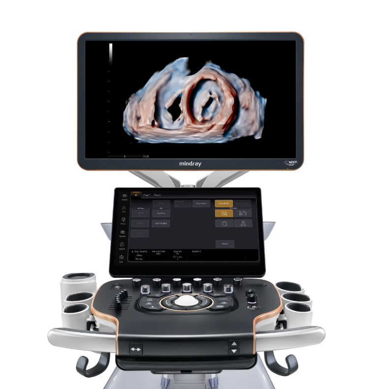
Ergonomics
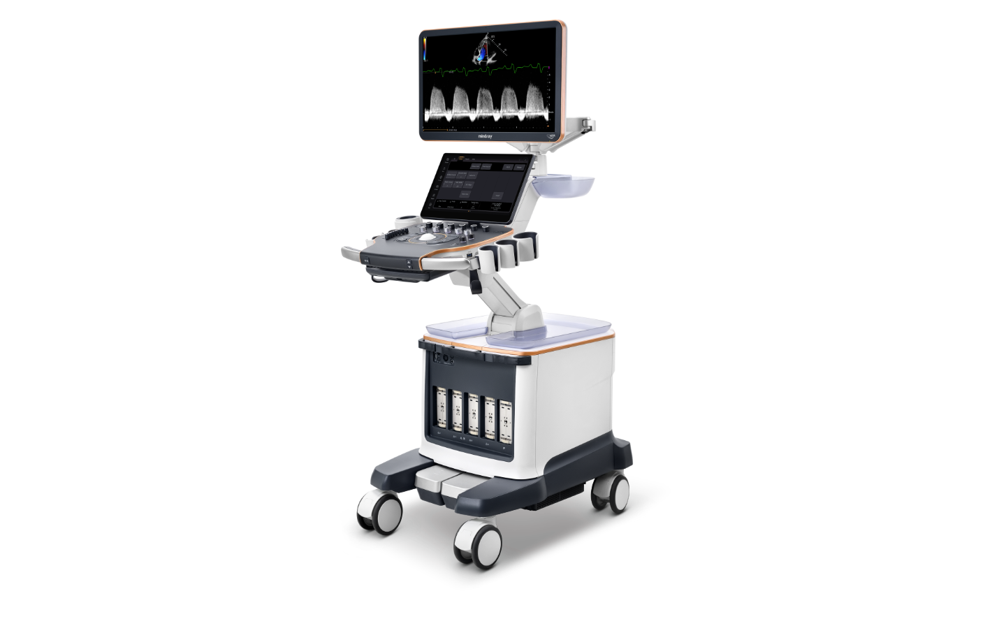

23.8' Ultra HD Diagnostic Display

15.6' High Sensitivity Touchscreen

Five transducer ports

Dedicated CV Control Panel


1
23.8' Ultra HD Diagnostic Display
2
15.6' High Sensitivity Touchscreen
3
Dedicated CV Control Panel
4
Five transducer ports

Cardiovascular Transducers Family
Cardiovascular transducers family covering cardiac examination items, including 3D TTE/TEE transducers, multi-dimensional single crystal transducers, linear transducers and so on.
Clinical Images

Normal A4C

Mitral and tricuspid valve regurgitation

Spectrum of aortic valve regurgitation

Posterior mitral valve prolapse

Mid-esophageal X-Vue for long axis view

3D TEE image of MV (surgical view)

Popliteal artery and vein

Carotid blood flow