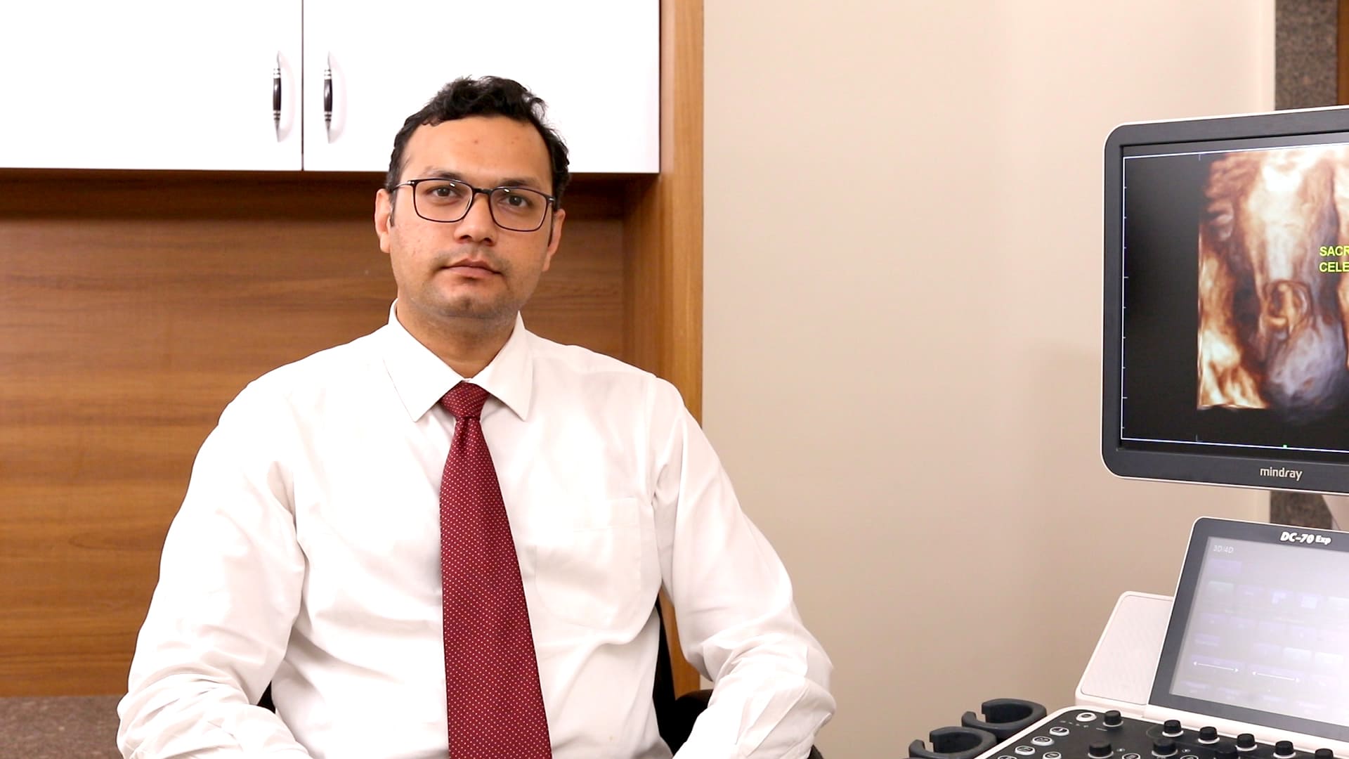
Dr. Devang Patel
Feto Maternal Medicine Specialist, Consultant at CIMS Hospital & Gujarat Fetal Medicine Centre, Ahmadabad
Dr Devang Patel is a renowned gynaecologist & Feto Maternal Medicine Specialist specializing in high-risk pregnancy and fetal medicine. He is a consultant at Care Institute of Medical Sciences (CIMS Hospital) and also attached with Gujarat Fetal Medicine Center, Ahmedabad
Dr Patel’s special area of interest includes invasive procedures like Amniocentesis , Chorionic Villus Sampling , Fetal reduction , Intra uterine blood transfusion, fetal echo, multifetal pregnancy, Aneuploidy screening. He also specialises in Hypertensive Disorders, Medical Disorders with Pregnancy like APLA , SLE , Jaundice and Thrombocytopenia in Pregnancy. Dr Patel is a specialist in management of Morbidly Adherent Placenta (PL Accreta) & Recurrent Pregnancy Loss.
Dr Patel talks about the emerging state of fetal imaging in the country and the benefits of modern imaging technology
Question 1: Can you comment on the current state of fetal imaging and prenatal care in our country?
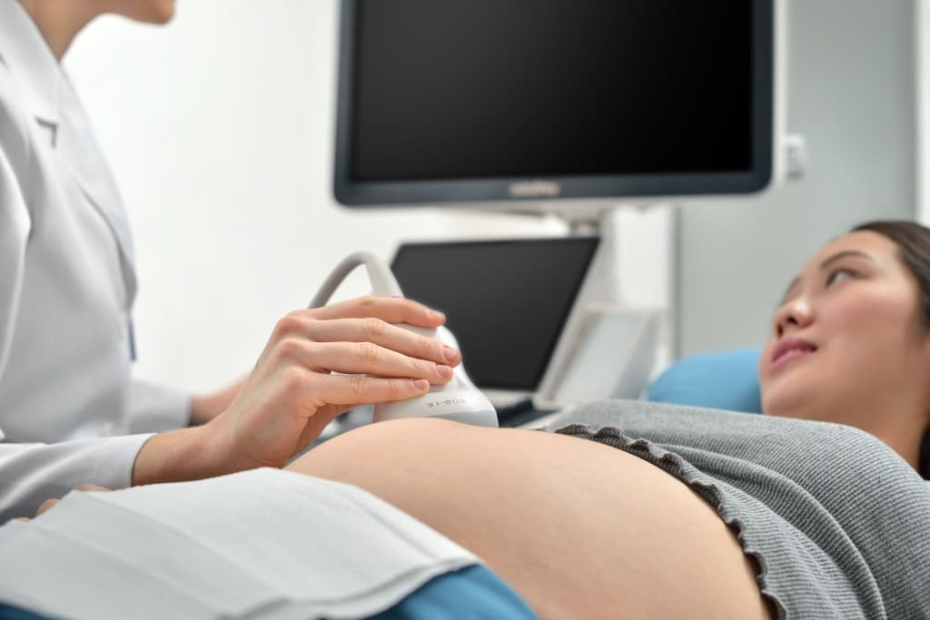
Answer: Prenatal ultrasound scenario in India is still finding its path to make an overall impact. Only 24 percent pregnant women are hardly undergoing one ultrasonography throughout their pregnancy. It means almost 3/4th of the pregnant women do not get the facility of the ultrasound. Primarily there is a lack of awareness about the importance of the imaging system during pregnancy. This is a cause of concern as prenatal ultrasound imaging helps us to diagnose several anomalies like neural tube defect and certain heart conditions in the fetus during antenatal period. However we are very confident this condition will improve soon.
Question 2: Which are the most commonly seen fetal abnormalities nowadays?
Answer: In our experience of antenatal ultrasound, the most common defects which are found are neural tube defect, heart conditions like ventricular septal defect, transposition of great arteries and also renal anomalies.
Question 3: What latest technology advances are observed in fetal imaging? How are modern fetal ultrasound technologies helping in diagnosis of the most complex fetal defects?
Answer: With the latest technology advancement in 3-D and 4-D ultrasound imaging, we are better equipped in diagnosis and prognostication. Secondly advanced technology facilitates easier communication with the patients as well as the referring consultant. For example, if we find a cleft lip in the fetus during imaging, we can present the actual 3D image to the patient and the referring doctor. It helps to convey the presence of such a condition clearly.
Question 4: Owing to advancements in 3D and 4D ultrasound technologies, how is advanced visualization helping in determining congenital defects much better than earlier?
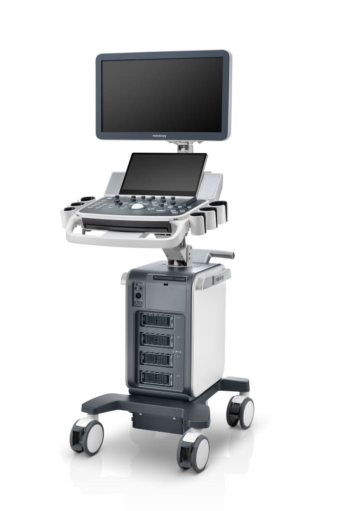
Answer: With the advancement in technology like 3-D and 4-D imaging, Colour Doppler, Power Doppler, Fetal STIC etc. from Mindray ultrasound, we get a clear diagnosis of several anomalies like that of the heart during the antenatal period. With these results, you can actually counsel the patients with a better approach and they can be prepared for any further corrective measure. If the condition is life threatening or the prognosis is poor, they have the option of termination of pregnancy. However, if the fetus has minor birth defects, they can be monitored well in the antenatal period and can be delivered at the tertiary perinatal Centre. Thus, the baby can be taken care of in the best supervision of experts. These are the advances in fetal echocardiography which has decreased the perinatal morbidity and mortality caused due to prenatal defects.
Question 5: what is the future scope and potential of growth of fetal imaging in our country?
Answer: There is a huge lacunae to be filled in the field of fetal medicine and antenatal ultrasound, especially in India. So, there is increased scope for the usage and development of this technology in India where we can provide these facilities to almost 3/4th of the antenatal mothers who cannot avail this technology at present. For more information or consultation, Dr Patel can be reached at : devangpatel05@gmail.com