Q: How would you describe the role of ultrasound in the field and its significance in the diagnosis and management of pelvic floor disorders?
D: Ultrasound is essential for certain procedures due to technical reasons. For instance, when it comes to imaging Gynecological Implants, Urogynecology Implants, slings, and measures, CT and MRI scans fall short in providing clear visibility. Ultrasound, on the other hand, excels in capturing these structures effectively. While this distinction may be obvious in some cases, it may not be as apparent in others. Cross-sectional imaging techniques like CT or MRI encounter limitations in the pelvis due to the presence of bone. CT scans, while beneficial for bone imaging, struggle to visualize soft tissue structures in the pelvic floor. In contrast, MRI offers superior resolution for soft tissues in a cross-sectional manner, particularly in the axial plane. That means ultrasound's technical advantages make it indispensable for addressing the need for improved accuracy in pelvic floor assessment.

Q: Are there any successful initiatives or innovative approaches that you have seen or been involved in, which have significantly improved attention to pelvic floor health with the use of ultrasound?
D: What I tried to establish from 2008 onwards was a service where women can access a pelvic floor assessment after childbirth, in particular in those who are 35 or older when they have their first baby. Difficult births involving large babies, anal or vaginal tears, and interventions like vacuum or forceps delivery can cause distortion. I believe it's crucial to offer women a pelvic floor assessment at 6, 8, 10, or 12 weeks after childbirth. This assessment aims to provide reassurance that everything is fine or identify unnoticed tears. We want to ask if they have noticed any tears and offer guidance for the future to prevent complications.
Similar to treating a sports injury, a pelvic floor physiotherapist can play a key role in addressing these muscular-skeletal injuries. They can guide women on muscle training and compensatory hypertrophy in surrounding muscles to improve strength. Raising public awareness is essential, and companies like Mindray have the means to contribute significantly to this objective.
Q: As an influential figure in the medical community, during your global education tour, can you share examples of successful collaborations and the impact these workshops and conference have had on healthcare outcomes?

D: Like a strawberry plant that sends out shoots, puts down roots, and produces more shoots, a similar pattern has emerged in my education journey. Since 2005, approximately 180 doctors have completed a standard five-week attachment in my unit. Each year, between 10 and 20 doctors came for training, with the number averaging around 15 in later years. Many of these doctors, much like the shoots of a strawberry plant, have gone on to train others in their local communities, such as in the Philippines.
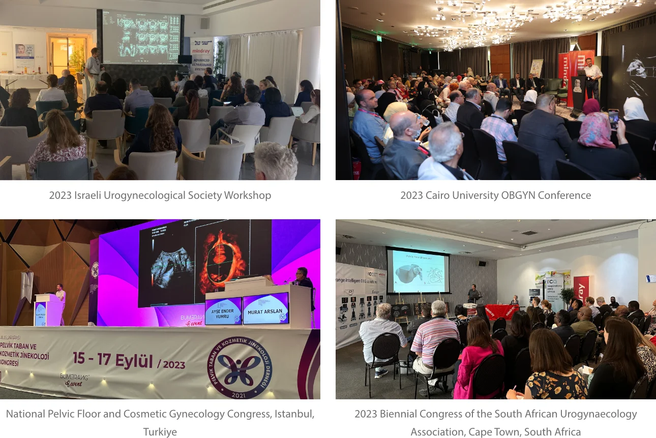
Recently, I had the privilege of being a keynote speaker and workshop facilitator in Manila, where a large group of around 800 attendees gathered. While it may not sound significant, this turnout was impressive for the Philippines and even for Australia. Among the attendees, I was pleased to see five doctors whom I had previously trained, and I realized that several of them have now established their groups and engaged in teaching activities. The impact of my work has extended to various countries, including Norway, Sweden, Denmark, the Netherlands, Indonesia, the Philippines, Thailand, and notably China. My unit has hosted around 12 to 15 doctors from China alone, showcasing the widespread adoption of the approaches and practices I have shared.
Q: During your visit to Mindray HQ, have you come across any specific initiatives or technologies that you believe have the potential to revolutionize the way doctors approach to urogynecology, or solve the challenges in childbirth- related pelvic floor trauma?
D: During my recent talks in Manila, Guangzhou, and Sydney, one fascinating topic I discussed was the application of artificial intelligence (AI) in image analysis. As early as 2008, we conducted a study on automated analysis, which has now proven to be highly beneficial. With advancements like Mindray's Smart Pelvic system, among other similar software, operators are assisted in documenting muscle distensibility, a crucial step in assessing pelvic floor damage.
At one of my clinics, we have been comparing the automated system with the standard manual process. While the manual process takes 2 minutes, the automated system completes the task in just one-tenth of a second. This offers substantial workflow optimization, faster results, and provides less experienced operators with guidance on desired outcomes.
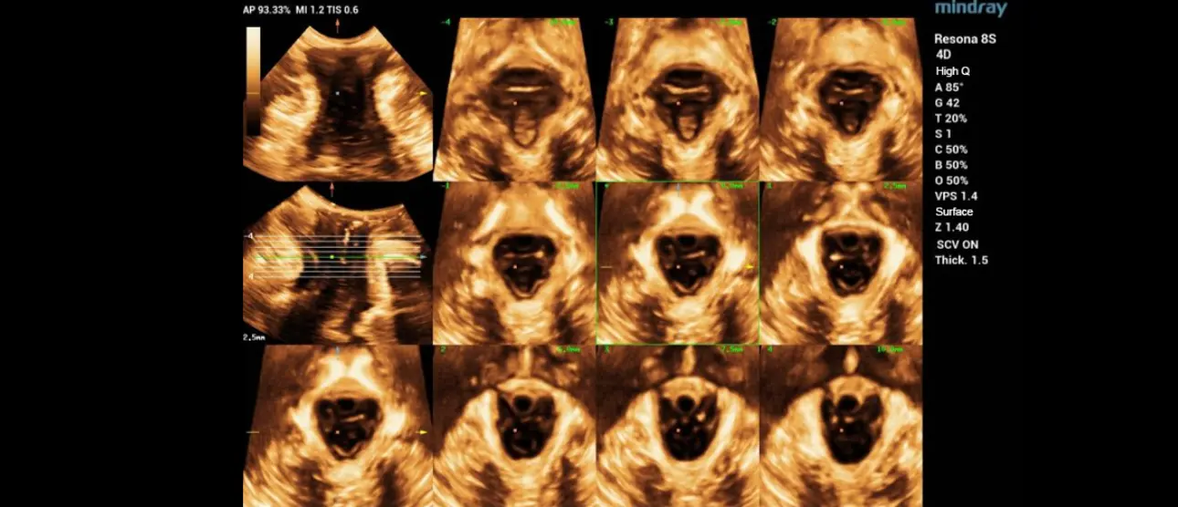
A significant development is Mindray's automated tomographic assessment of the anal sphincter, a first-of-its-kind innovation. In approximately 5% of women experiencing their first childbirth, the anal sphincter can sustain severe damage. Traditionally, obstetricians are familiar with suturing such tears. Now, the integrated software in these systems produces a standardized set of tomographic images with a simple push of a button, taking mere seconds. While some improvements are still needed, this progress marks just the beginning of exciting advancements in this field.

Q: As a mentor and educator, what advice would you offer to young doctors and healthcare professionals seeking to engage in meaningful collaborations with industry partners for the advancement of medical science and patient care?
D: Keep an open mind. Don't blindly trust everything stated in textbooks or what you observe around you. Constantly question established opinions, even those from highly regarded sources like Gray's Anatomy. As someone involved in revising the female pelvic floor chapter of this atlas, I discovered that around 6 to 7 out of the 15 to 20 illustrations were incorrect. We are currently rectifying these errors. This situation highlights the problem when even anatomical atlases contain inaccuracies. These are the foundational materials from which students learn. Instead, believe in your own truth based on your personal experiences. It is important to have mentors and trusted senior doctors, but it is not necessary to accept everything they say without question. Trust your instincts and knowledge while also being open to learning and guidance from others.

