


Nuewa R9 Platinum Edition
Inspiring Women's Healthcare
Inspiring Women's Healthcare
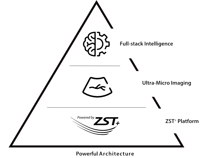
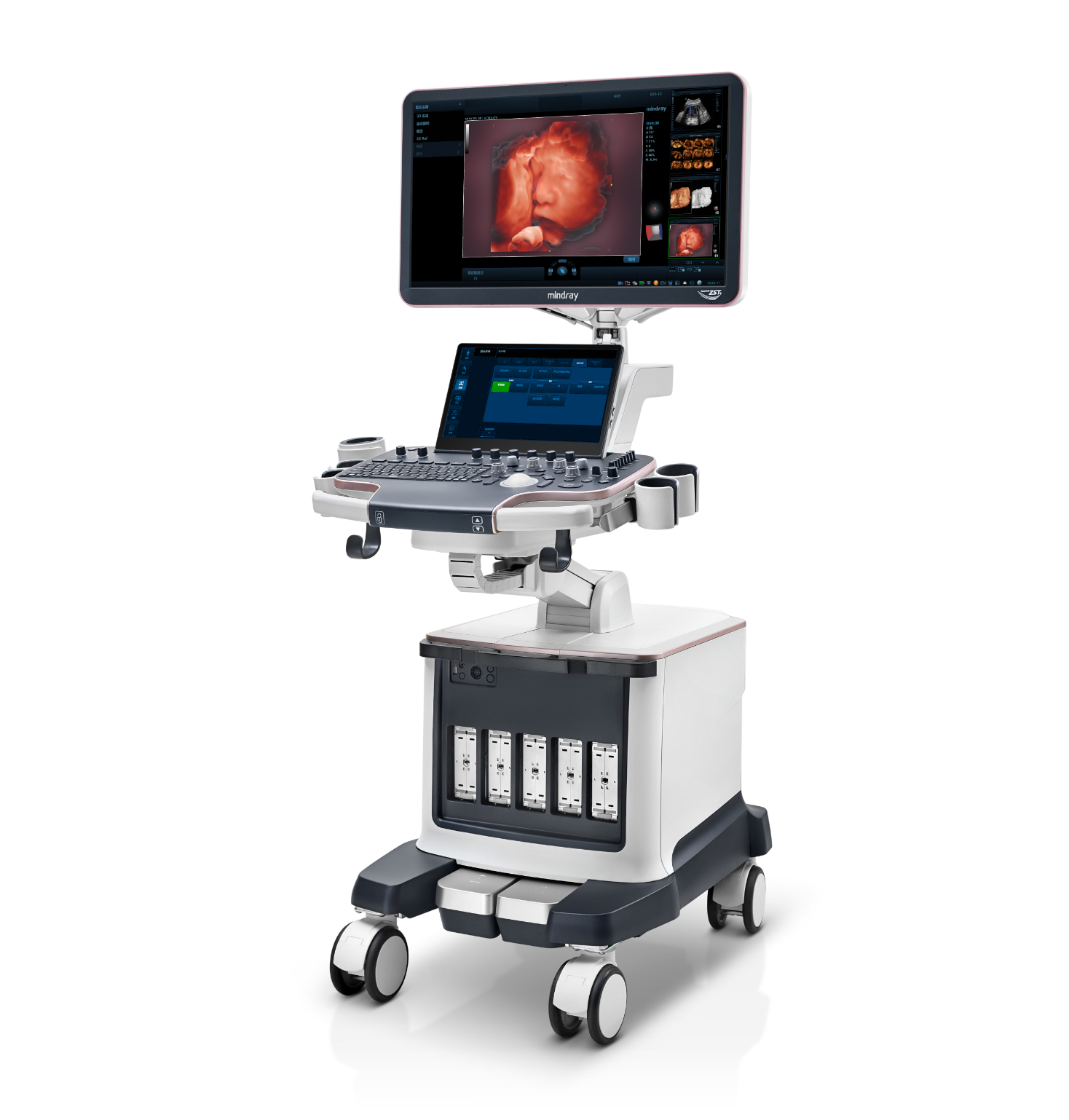
Thanks to the revolutionary ZST+ platform, Nuewa R9 is powered by evolving leading-edge technologies to meet diagnostic challenges in women's healthcare. Breakthrough technologies such as Ultra-Micro Angiography help you explore more micro-details and the truth beneath! Designed with the invaluable wisdom of experts, it innovatively provides full-stack intelligence beyond your expectation for diagnosis with more confidence, standardization and efficiency across different challenges in demanding and overburdened hospital settings.
Explore More Details and the Truth Beneath
UMA (Ultra-Micro Angiography)
The innovative technology breaks the bottlenecks of traditional Doppler imaging. With ultra-high spatial resolution and flow sensitivity, it allows detecting super-subtle and super-slow flow perfusions, thereby extending the clinical application of qualitative and quantitative ultrasound evaluation in fetal brain, kidney, placenta, endometrium, ovary, etc.
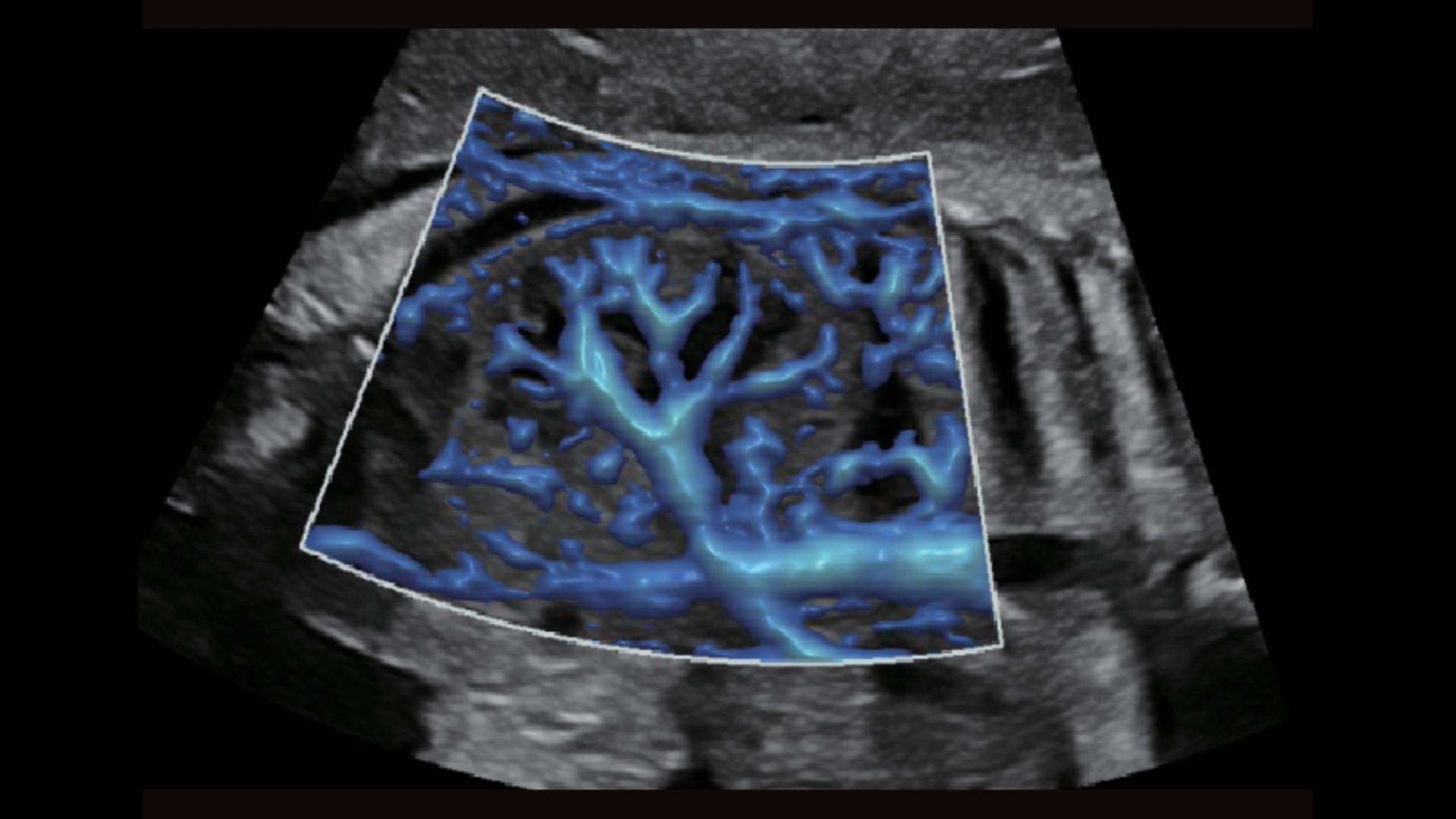
UMA - fetus renal flow
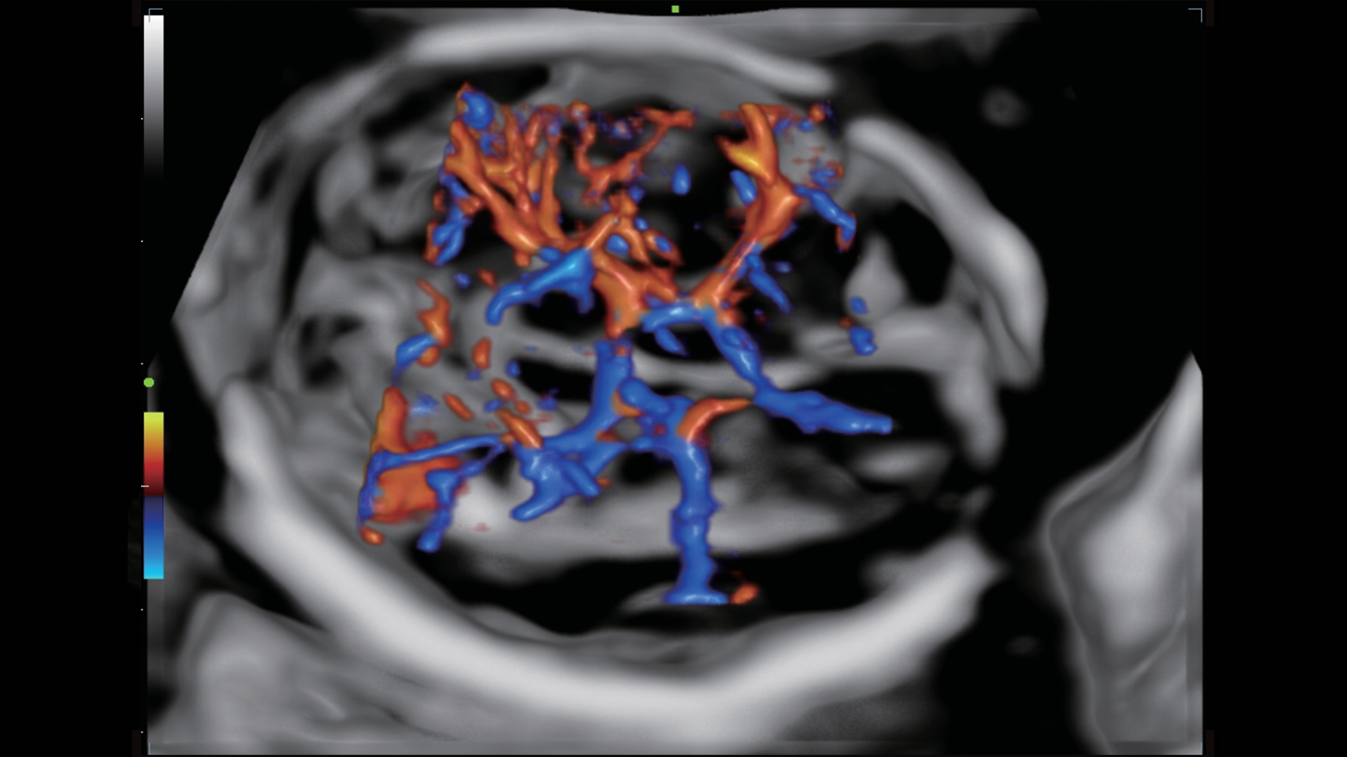
3D UMA - fetus intracranial flow
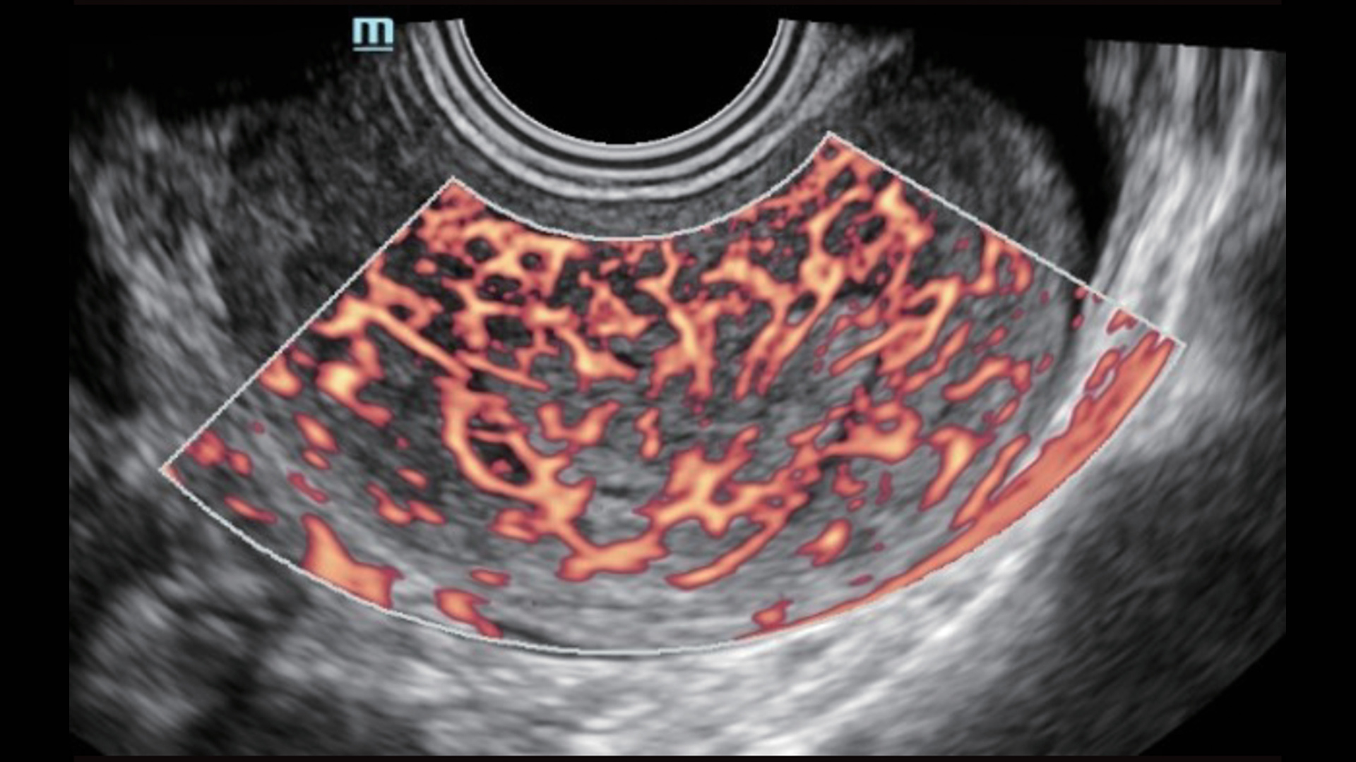
UMA - uterine and endometrial flow
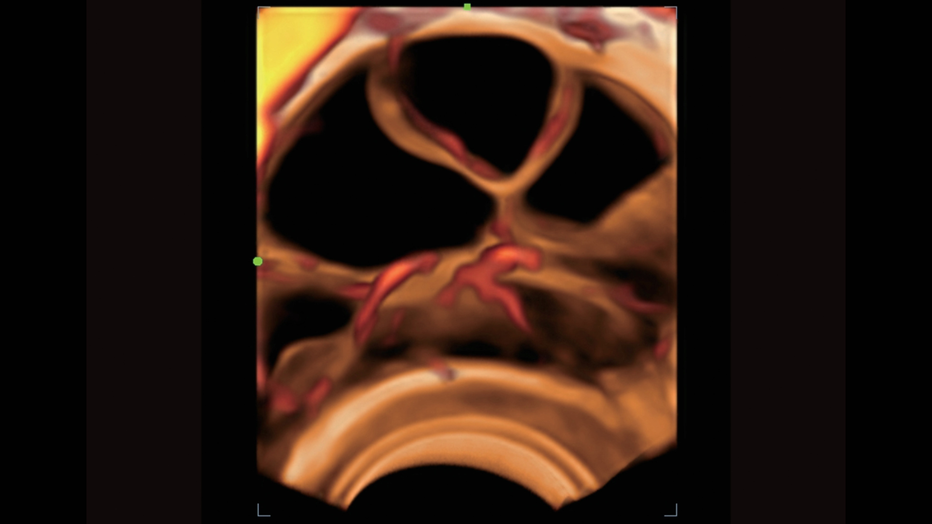
3D UMA - perifollicular blood flow
Scenario-oriented Full-stack Intelligence
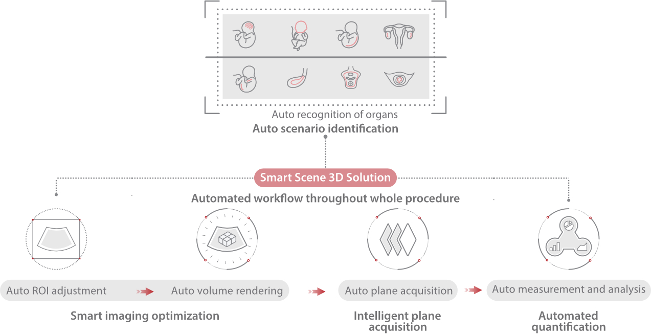
Combining advanced algorithms and domain-specific knowledge, the innovative Smart Scene solution enables auto identification of tissue characteristics and provides organ-specific diagnosis with full-stack intelligence. Based on auto scenario identification, the solution not only realizes smart 2D scanning with auto settings and measurements, but also delivers 3D full-stack intelligence in every step from volume imaging optimization to the difficult 2D plane acquisition, and quantification throughout whole procedure. It helps a lot in reducing the dependency on clinical skills, while increasing diagnostic accuracy, confidence and efficiency.
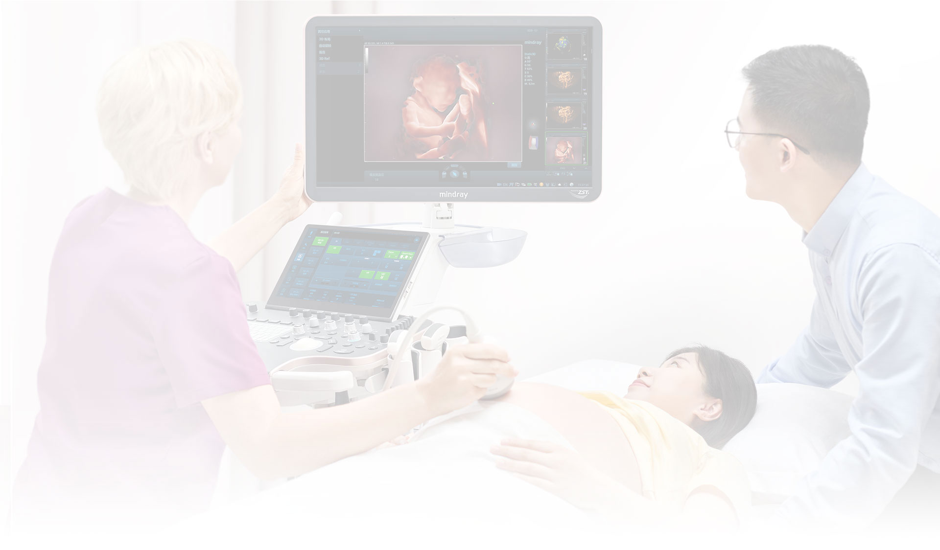
Professional Ultrasound Solutions with Full-stack Intelligence
- Protecting the new lives with fertility and obstetrics solutions
- Caring for women's health with gynecology and urogynecology solutions





 Fertility
Ovarian Reserve Assessment
The brand new Smart FLC provides precise volume rendering, count and quantitative analysis of ovary and even antral follicles for easy, effective and confident ovarian reserve assessment.
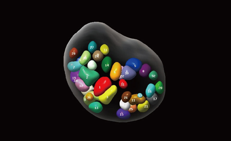
Precise imaging of ovary and follicles
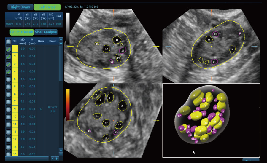
Intelligent follicle grouping and count
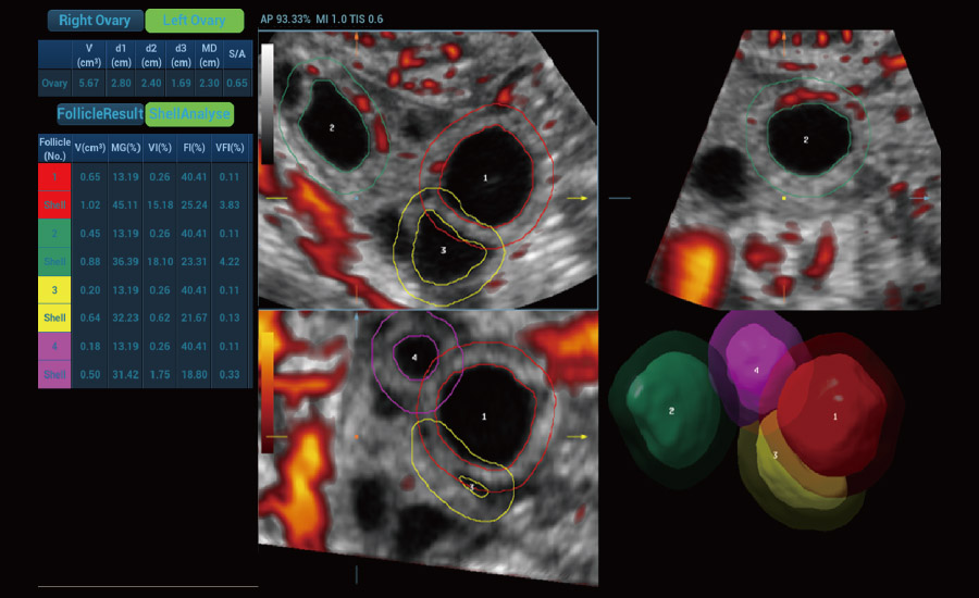
3D analysis with shell for perifollicular flow
Endometrial Receptivity Analysis
Smart scene-endometrium realizes intelligent endometrial thickness measurement, volume rendering, auto coronal plane acquisition, and even 2D or 3D vascularity study for endometrial receptivity analysis.
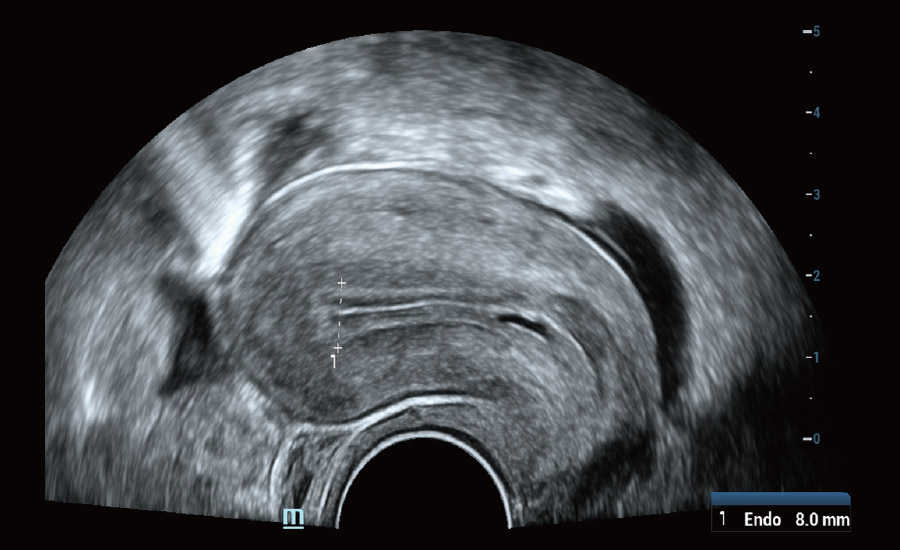
Auto measurement of endometrial thickness
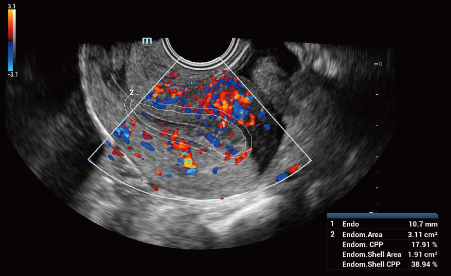
Auto tracing and flow analysis of endometrium
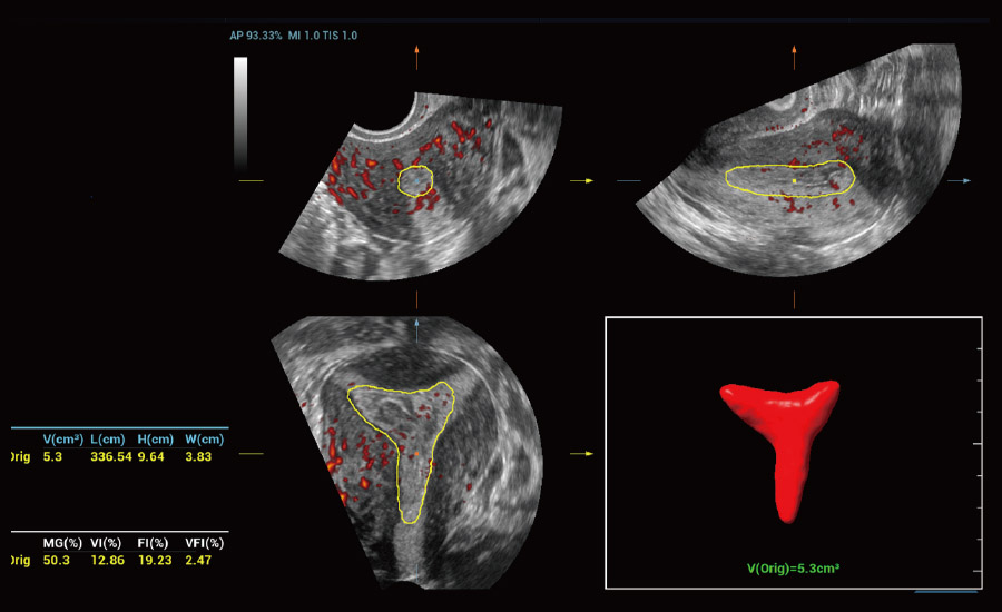
Auto volume rendering and quantification
Fallopian Tube Patency Evaluation
Easily and intuitively explore the tubal patency and direct the type of
treatment
that is required for women having trouble falling pregnant.
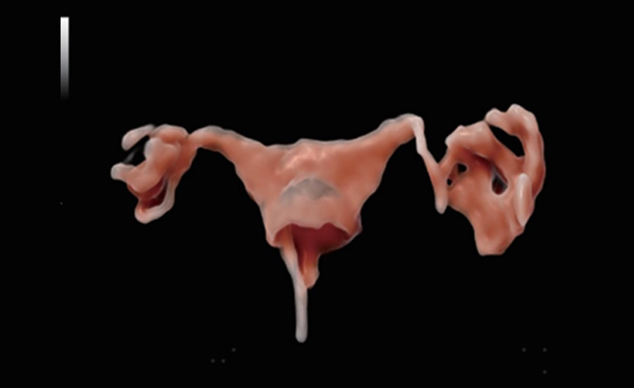
4D HyCoSy with iLive
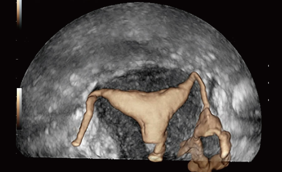
4D Tissue-Contrast Mix Rendering
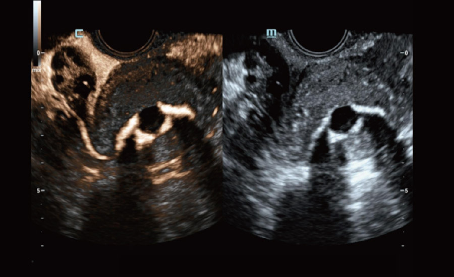
2D Tissue-Contrast Mix Rendering
 Obstetrics
Excellent Imaging for Prenatal Diagnosis

Early OB NT

Fetal hydronephrosis
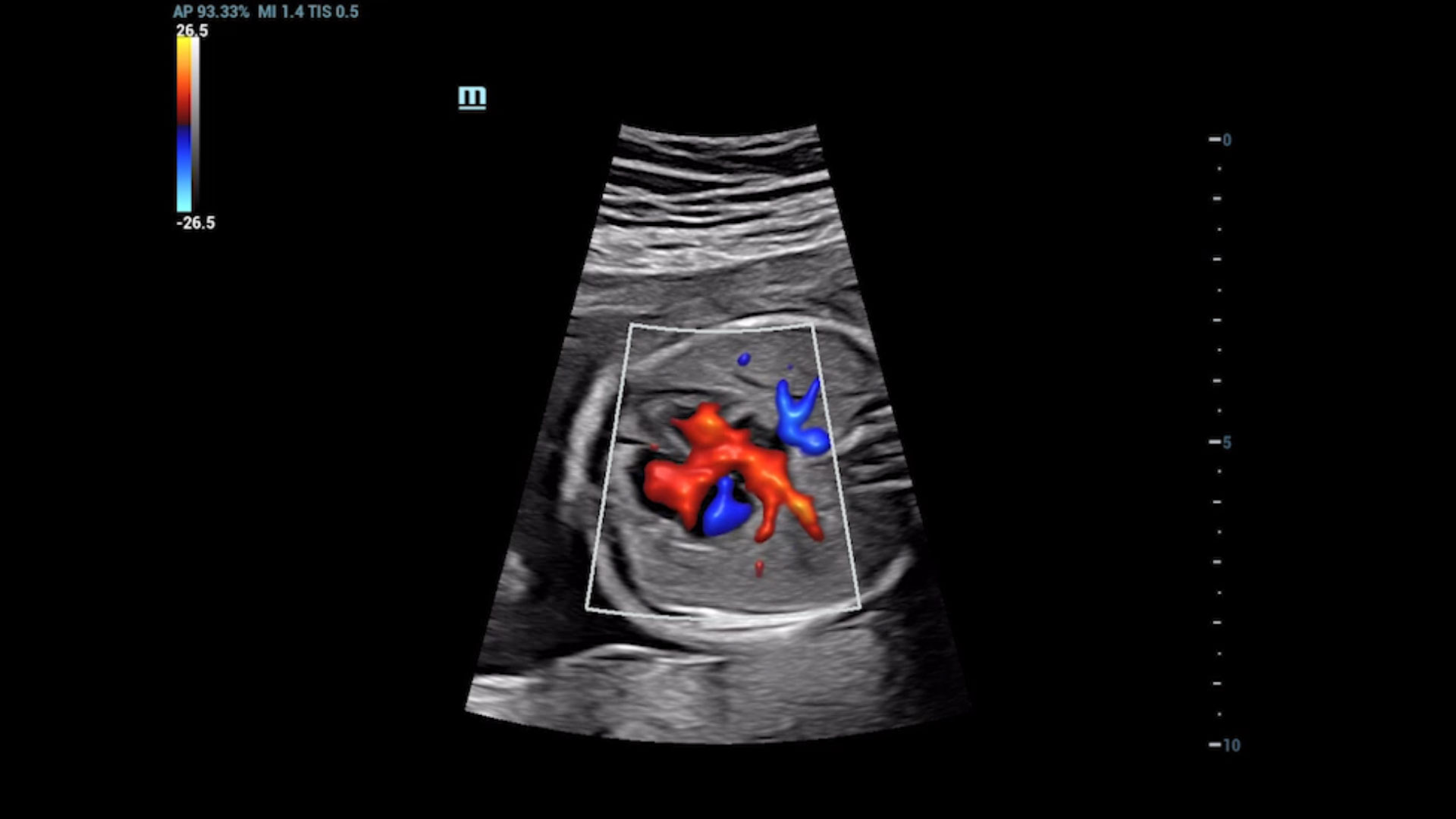
4-chamber Color flow
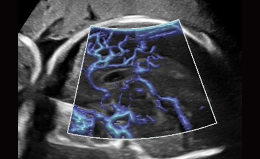
Pericallosal flow - UMA
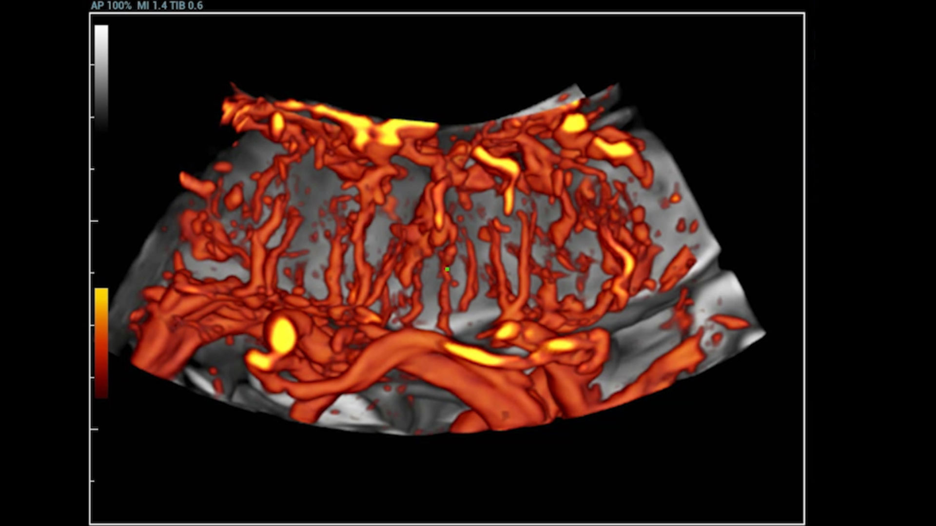
Placental flow perfusion - UMA 3D
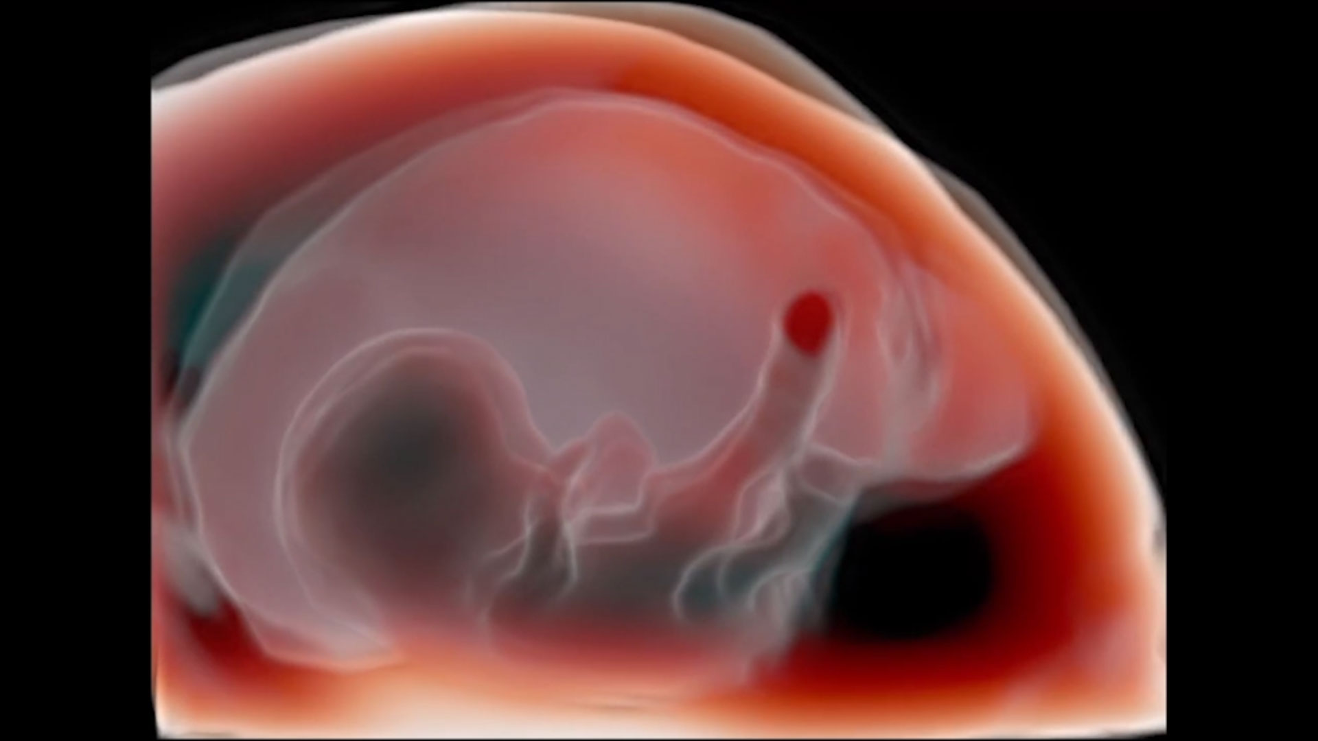
Early OB - iLIve
Smart and Accurate CNS Diagnosis - Fetal Brain
High-clarity images with subtle details and Smart Scene intelligence help to easily acquire the most significant planes and obtain frequently used measurements of fetal brain for smart diagnosis, enabling improved throughput and reduced user dependency.
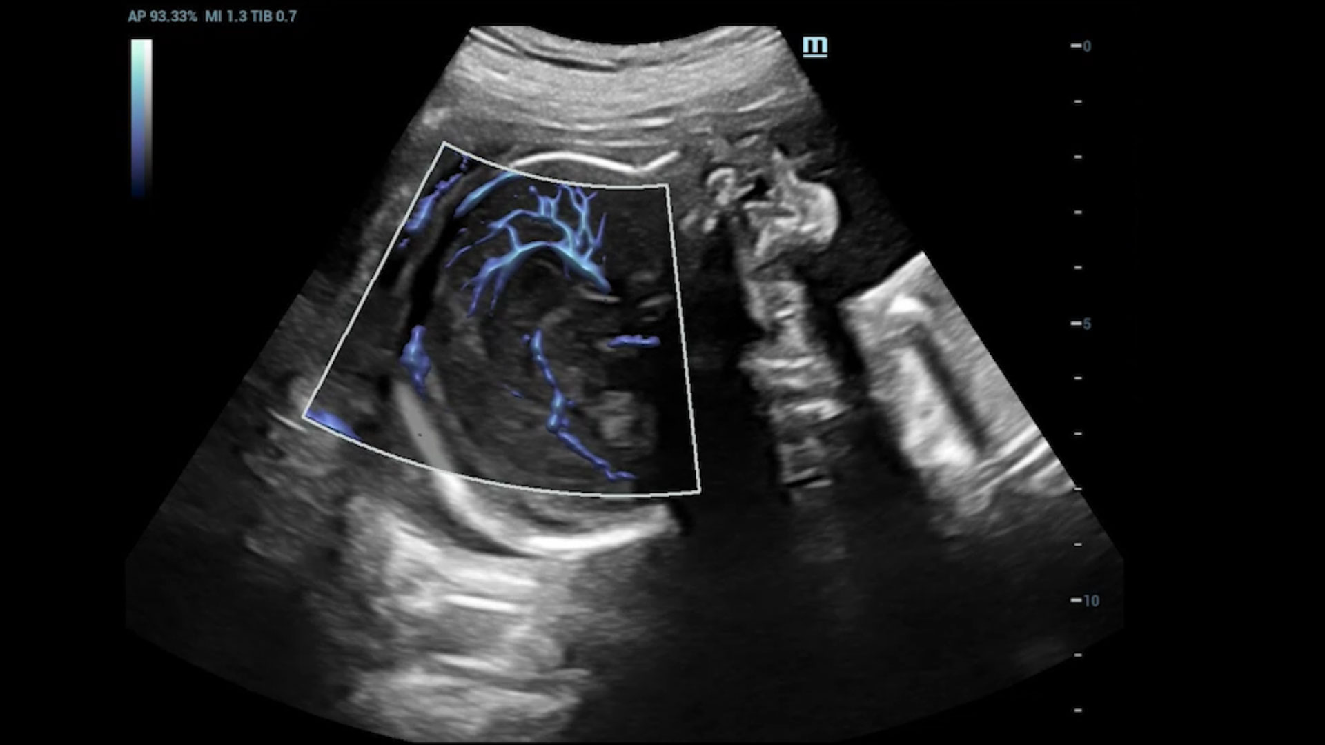
Pericallosal flow - UMA

Smart Planes CNS
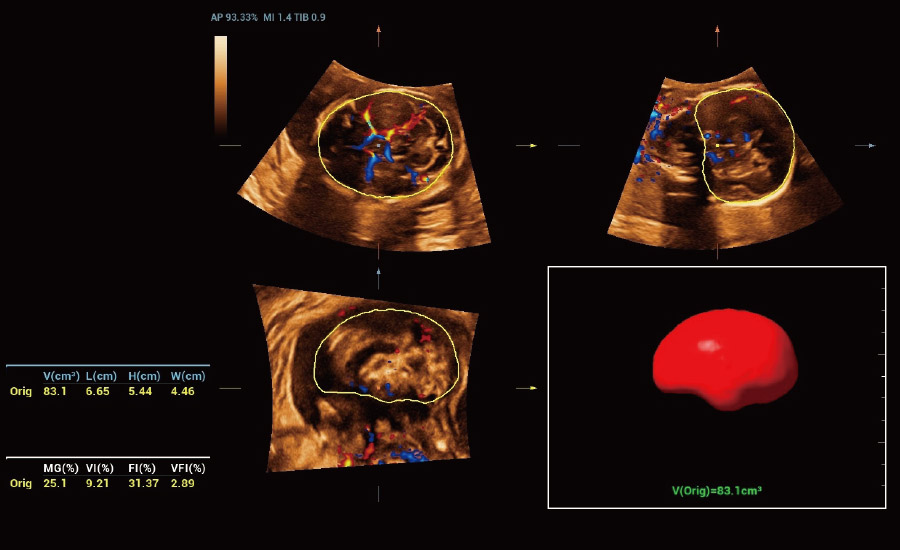
Smart ICV with UMA 3D
Easy and Smart CNS Diagnosis - Fetal Spine
Smart Scene 3D-spine reduces the difficulty in scanning and intelligently splits vertebral bodies and arches for clear observation and reduced misdiagnosis of fetal spina bifida. The guide line also enables much easier and more confident positioning of conus medullaris.
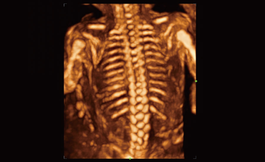
Fetal spine - regular 3D

Auto splitting of vertebral bodies and arches
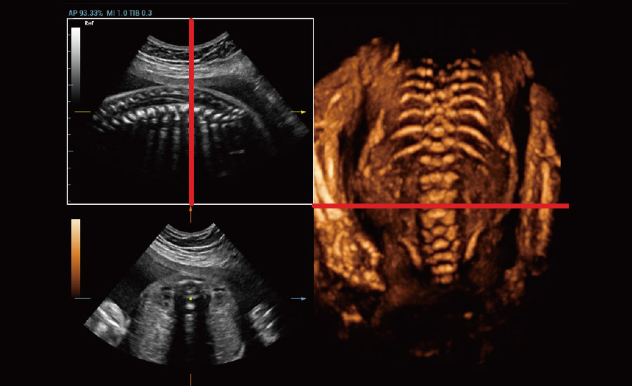
Easy positioning of fetal conus medullaris
Precise Fetal Heart Evaluation and Study
Excellent fetal cardiac 2D and flow imaging ensures diagnostic confidence. A combination of tools enhances the efficiency and provides more information, such as auto monitoring of fetal heart rates, accurate evaluation of fetal heart function, and myocardium motion study.
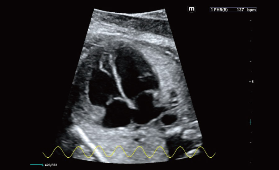
Smart Fetal HR
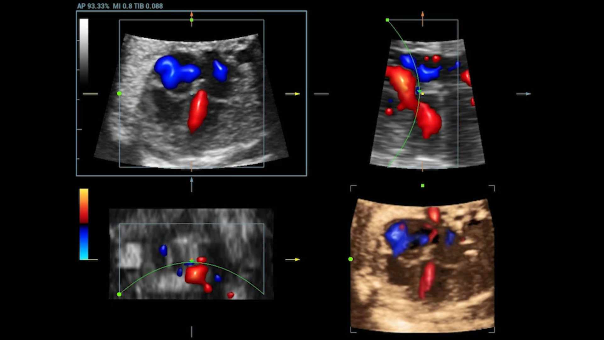
Color STIC
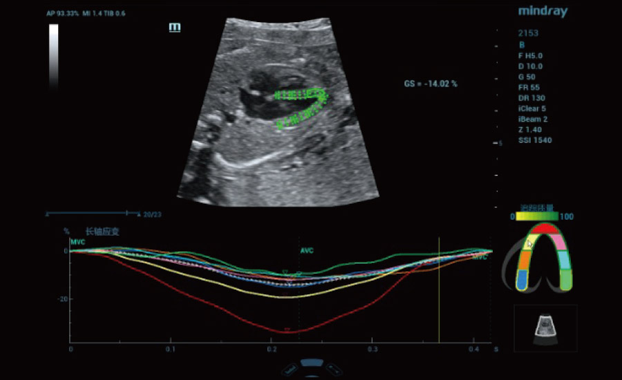
Fetal heart TT QA
 Gynecology
Smart, Effective and Efficient GYN Scan
Ultrasound plays a pivotal role not only in the regular examination of uterine, ovarian and adnexa, but also in the detection and characterization of abnormalities. Effective and efficient scan is enabled by a series of smart tools.
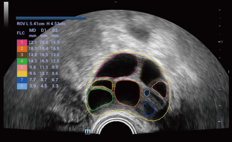
Auto measurement of ovary and follicles
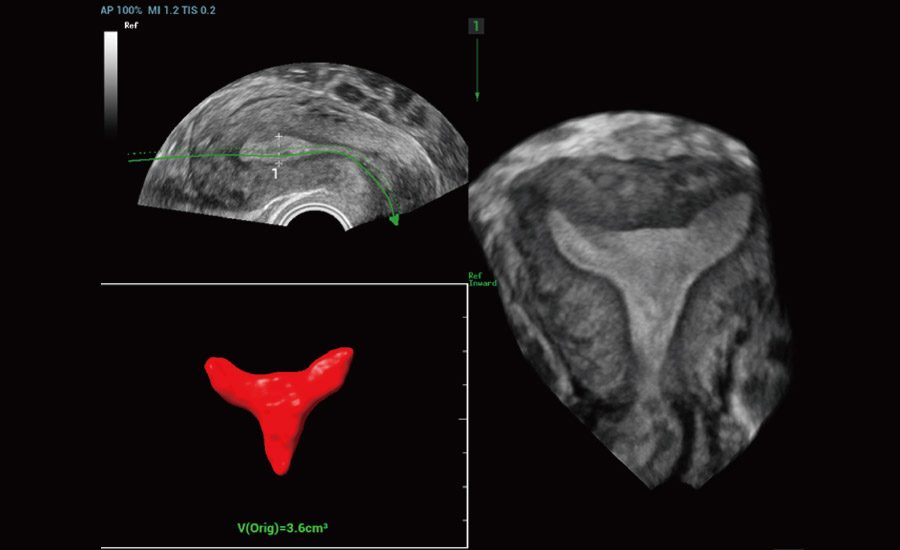
Auto imaging of endometrium coronal plane
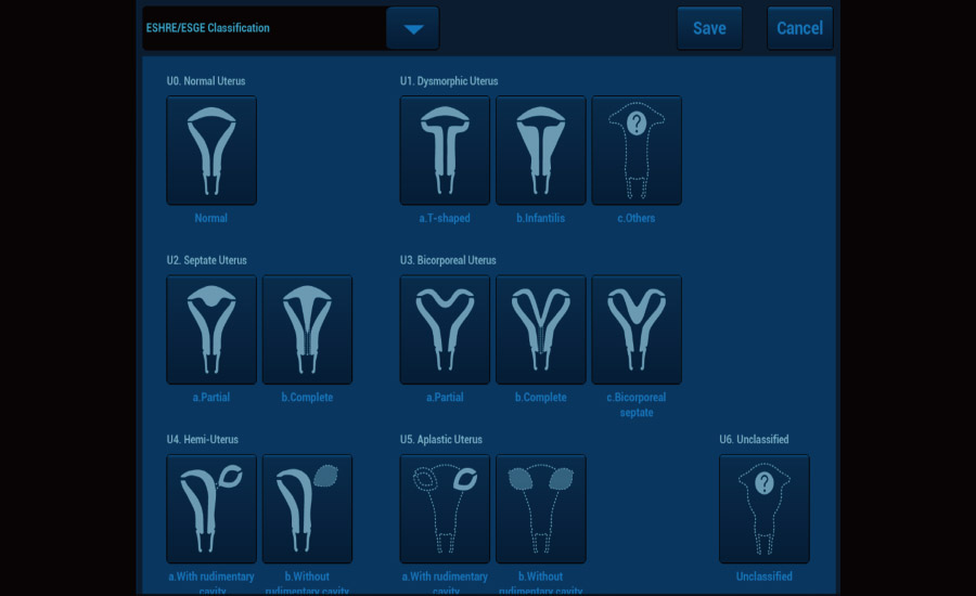
Guidance for uterine malformation classification
Multi-modal Imaging for Lesion Detection and Characterization
Multiple imaging modes and analysis tools allow comprehensive evaluation of the lesion, including 2D/3D tissue structure, hemodynamics, micro-flow perfusion, tissue stiffness, and quantification, giving more confidence for diagnosis, especially of complex GYN tumors.
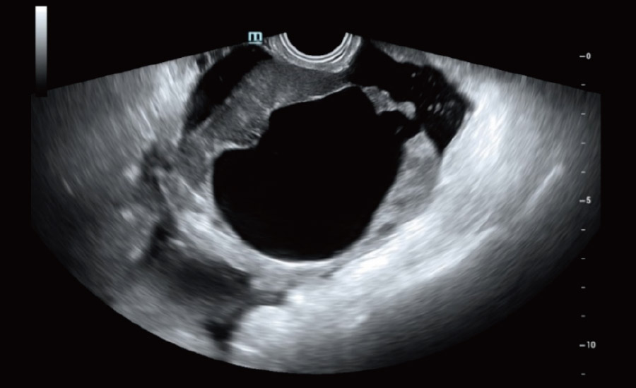
Ovary lesion - 2D
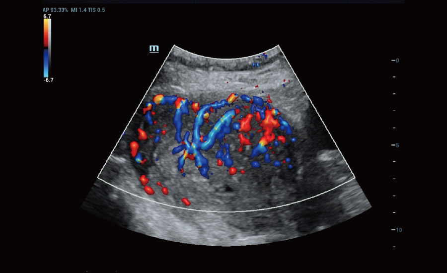
GYN mass - Color
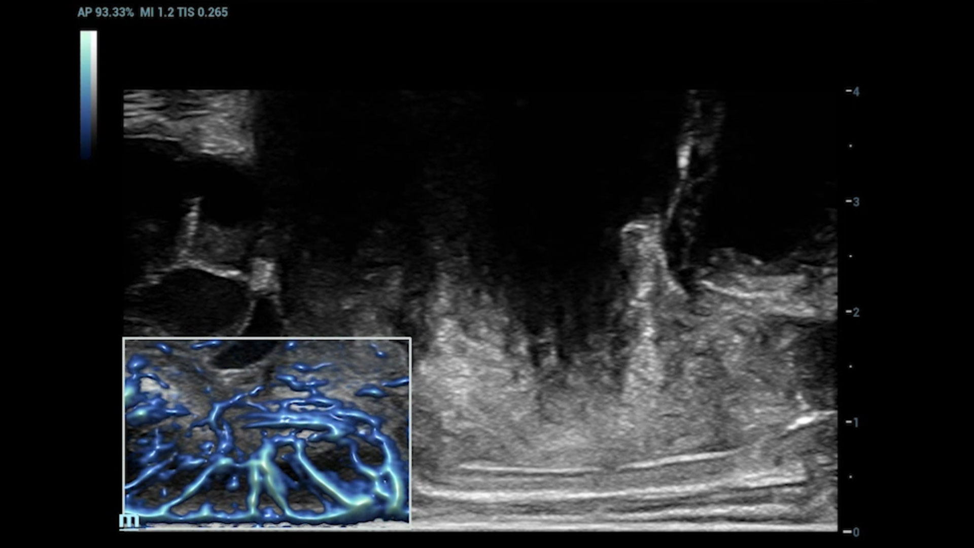
Endometriosis - UMA
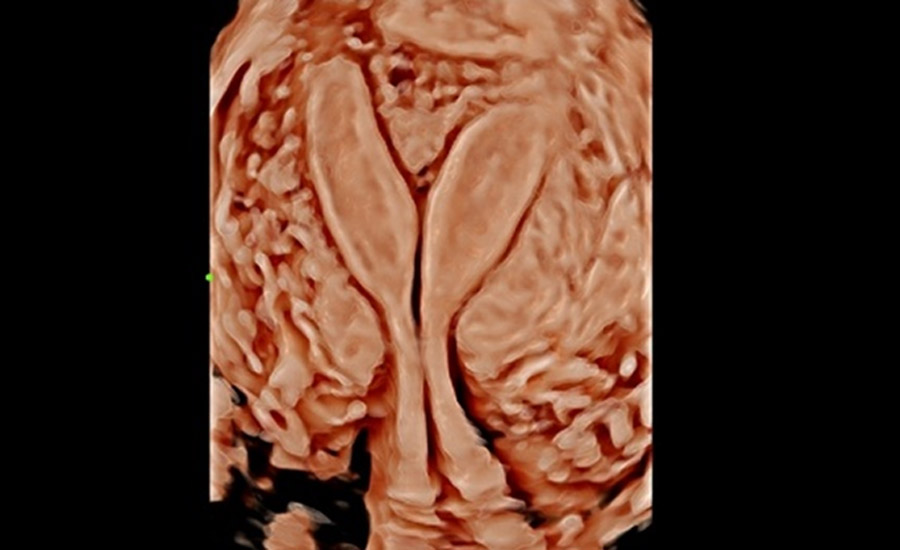
Mediastinum uterus - 3D
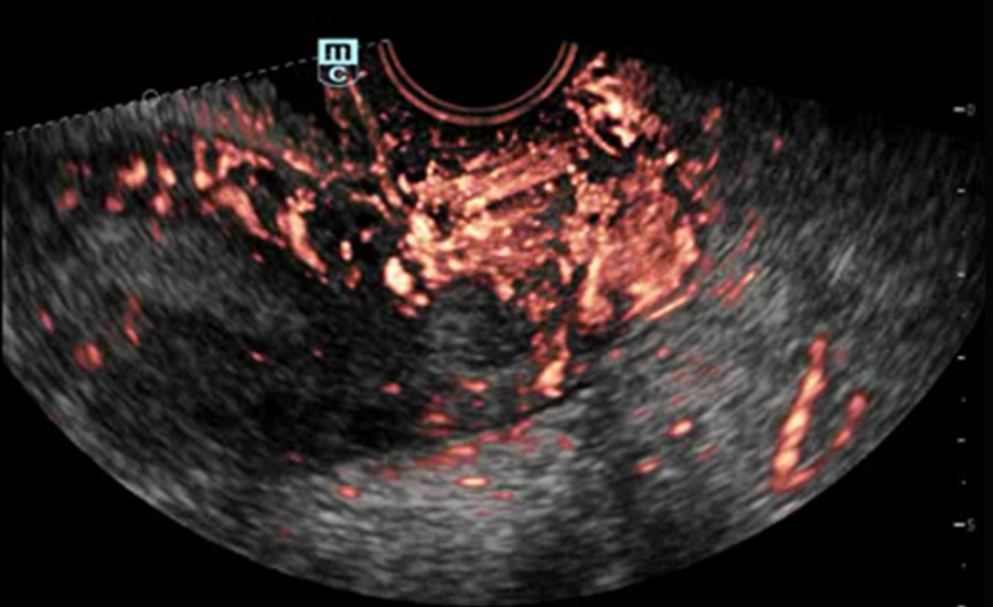
Cervical cancer - CEUS (tissue & contrast mix)
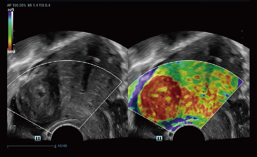
Uterine myoma - Elastography
 Urogynecology
Smart Assessment of Mid-saggital Pelvic Scan
With a simplified user interface, it generates a standard coordinate system and automatically provides all related measurements within seconds.
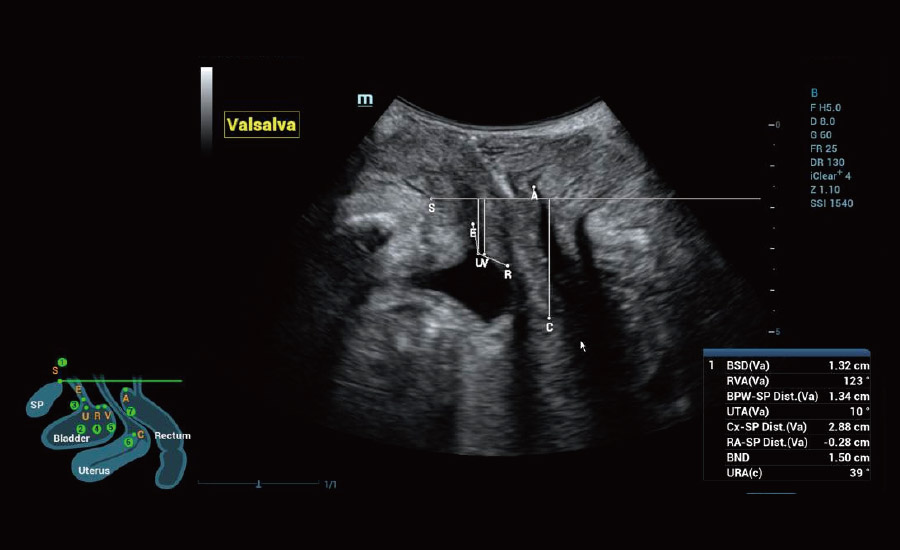
2D auto evaluation - Smart Pelvic
Intelligent Analysis of Levator Hiatus
It provides full-stack intelligence for volume imaging, the ability to acquire multiple planes, and automated accurate measurements to enable easy evaluation of levator ani muscles.
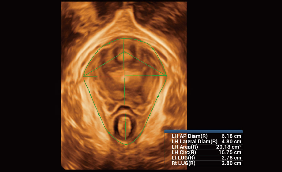
Auto volume imaging and measurement
Auto Acquisition of Anal Sphincter Planes
Smart Scene 3D can automatically detect anal canal scanning Smart Scene 3D can automatically detect anal canal scanning sphincter for easy evaluation of its integrity and continuity.
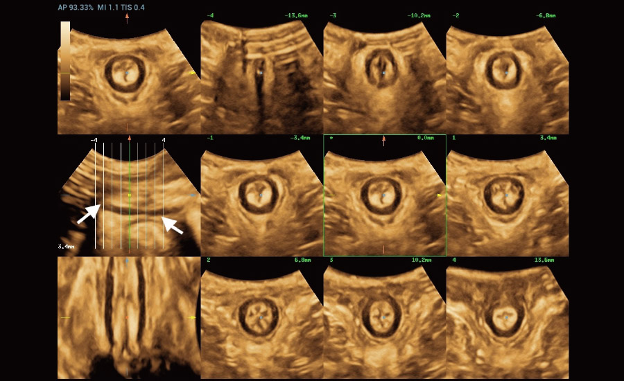
Auto acquisition of anal sphincter planes

Ensuring a Better User Experience
Nuewa R9 is innovatively designed with end users in mind. It features a 23.8" HD monitor, gesture control touch screen, dual-wing floating arm and a six-way floating control panel to enable an agile, smart and intuitive user experience beyond your expectations.
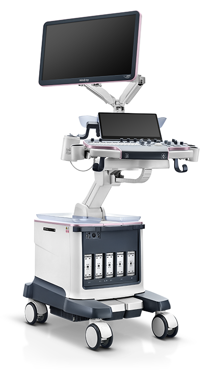
23.8" HD monitor
Rich imaging details
Auto brightness adjustment
Clear images even with light on
Dual-wing floating arm
More flexible monitor position
Much lower height during transportation
Stand-by batteries
Stand-by mode for up to 24H
6-way floating control panel
Up/down, back/forth, left/right
Electronic height adjustment
123.8" HD monitor
Rich imaging details
Auto brightness adjustment
Clear images even with light on
2Dual-wing floating arm
More flexible monitor position
Much lower height during transportation
3Stand-by batteries
Stand-by mode for up to 24H
46-way floating control panel
Up/down, back/forth, left/right
Electronic height adjustment