

Ultrasound today is the new stethoscope
Dr. Chander Lulla 2024-03-29
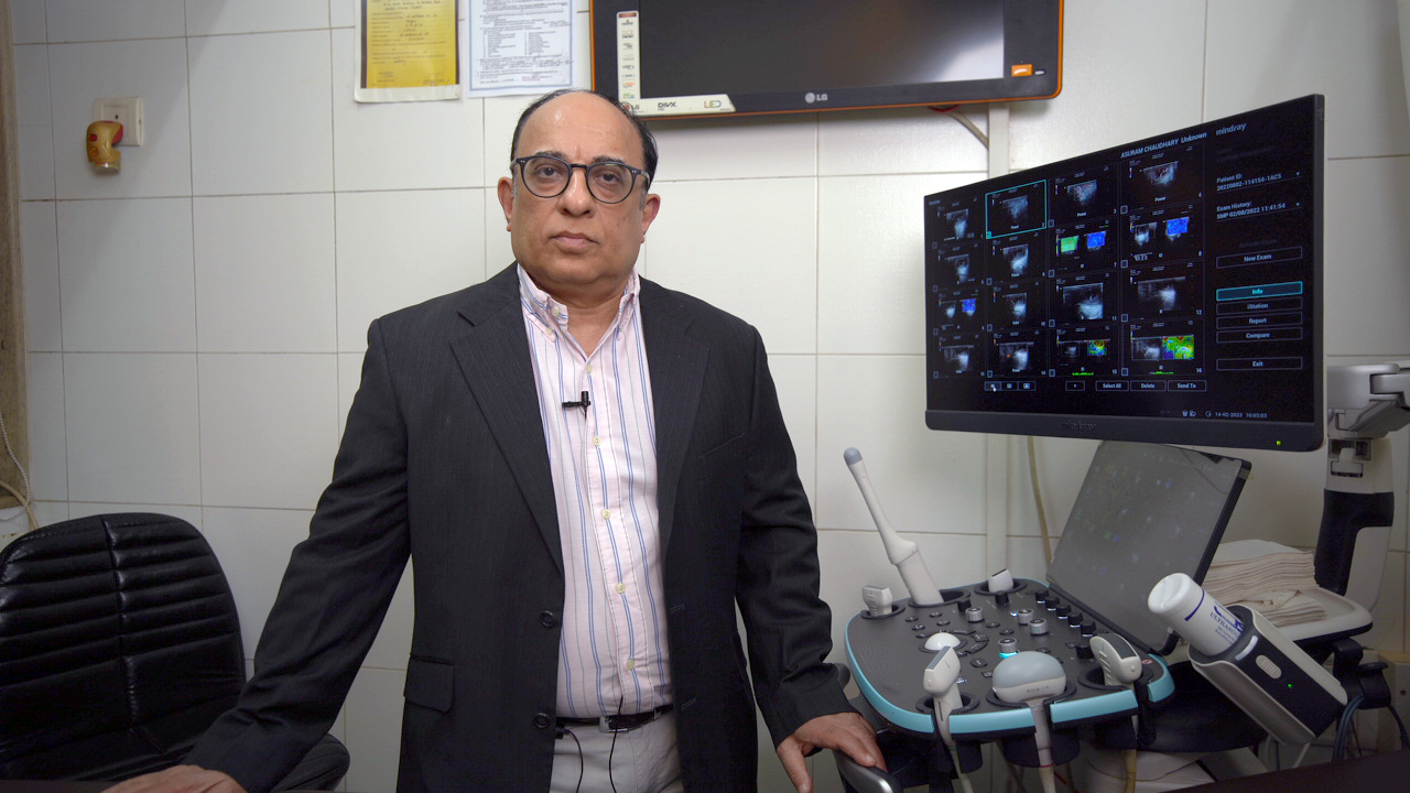
Dr. Chander Lulla
RIA Clinic, Mumbai
Dr. Chander Lulla is one of the most prominent radiologists in Mumbai and has more than 35 years of experience in this field. Dr. Chander Lulla practices at Ria-Clinic in Gamdevi, Mumbai. He completed his MBBS from the University Of Bombay in 1984 and MD - Radio Diagnosis/Radiology from the University Of Bombay in 1988. He also completed several trainings including color doppler USG from Guys Hospital, UK, Foetal Interventions from Kings College, UK, 3D Ultrasonography in 1999 from Seoul, Korea, 3D VISUS course from Vienna, Austria, and Foetal Medicine Foundation course from Sydney, Australia.
In this interview with Mindray India, Dr. Chander Lulla discusses the evolution of the ultrasound field, his experience with Resona I9 ultrasound system and much more
1. Mindray: You have more than 3 decades of experience as a radiologist. Can you please share the technological revolutions in the ultrasound field that you have witnessed?
Dr. Chander Lulla: In the last three decades of my experience, I have seen almost mind-boggling changes in ultrasound.
We started with basic portable small ultrasound machines, which used to be carried from one place to the other in the year 1987. Then, the mainframe machines were launched in the 1990s with colour, and doppler. It was one of the biggest game changers in ultrasound filed as blood flow was visible without placing the needle and injecting contrast. When I showed my first images of colour doppler at a conference during that period, people couldn't believe that we can see blood flow without doing an angiography. There were a lot of questions raised regarding the efficacy of the technology vis-a-vis angiography and others. Today, we can see that no ultrasound machine is complete without a stellar doppler component. Then in the year 1998, 3-D ultrasound created a revolution wherein we could see the baby’s face and the entire foetus in three dimensions.
Apart from that, we had multiplanar imaging that allowed us a great spatial orientation of different pathologies in the foetus or in the uterus. We could plan our interventive procedures or other procedures in a multiplanar format or in any plane instead of X, Y, and Z planes, we could rotate the volume into any plane.
Then the other significant development was, of course, elastography, which has been there from time immemorial. But with 2D shear wave elastography, we could do simultaneous imaging of the pathology and get shear wave measurements. So, this was very targeted, focused, and accurate, and we had a lot of quality checks that were possible.
2. Mindray: Which features of Resona I9 have helped you in your practice?
Dr. Chander Lulla: Resona I9 has been helping me in the clinical management of my patients.
i. Resolution
The most important thing for any ultrasound solution machine is the resolution and Resona I9 provides excellent and rapid spatial and contrast resolution. It doesn’t require focus zones and grants a complete harmonic and uniform image from the skin to a great depth.
We had a patient with a mass in his tongue and we performed sonography of the tongue using Resona I9. Because of the high resolution of Resona I9, we could see flow in the patient’s mass lesions because of microvascular imaging. The advanced elastography techniques using Resona I9 revealed that it could be a cystic mass and that the lesions had a small nodule. This degree of resolution was only possible due to the 23-megahertz transducer. Thanks to this new technology and high resolution, we can now obtain the same resolution for patients with high BMI as for patients with less BMI.
Without ResonaI9, patients would need a CT scan or MRI for more clarity.
ii. Ergonomics
Musculoskeletal problems are very common in radiologists due to the high-volume work and ergonomics. Resona I9 has been designed keeping ergonomics in mind and has helped me easily perform interventional procedures. It has a full-space floating control panel that can be rotated in a 360-degree fashion. The transducer connections are on the posterior side of the panel making it easy to be changed and the bottle warmer helps to make the patients feel more comfortable during the scan.
iii. Artificial intelligence
The artificial intelligence incorporated in Resona I9 enhances the workflow to a great extent and allows me to attend to my patients faster. Apart from that, it has innovative methods of calculation which is astounding as manual calculations are no longer required. For example, we can acquire the rapid volume of the brain in the foetus with ventriculomegaly. With just one touch of a button, we can get all the different planes and the measurements appear automatically. The BP, the head circumference around the cerebellar diameter, lateral ventricle diameter, brain volume, etc. can be seen very accurately.
I have been practicing for the past 30 years and I have gone through all the changes in ultrasound from Basic B mode ultrasound in the 1990s. Then we started doing colour Doppler in 91, 3D technology in 98, and Elastography in 2010. And now we have excellent AI-based technology where I don't have to physically measure so many parameters and even the basic parameters as it is all done by the machine.
So, it can be used for follicle monitoring, dynamic endometrial volume monitoring, and prostate monitoring on the volume transducer. We also have the biplane transrectal transducer, which is extremely beneficial for visualizing the prostate in two planes and allows me to do a great fusion biopsy. Apart from that, we have something called tissue tracking quantitative analysis for foetal echocardiography, where it can automatically track the endometrium and give us a functional analysis of the foetal heart in terms of ejection fraction, and the fractional lung volume, fractional volumes of the different chambers of the heart, and the cardiac output. Smart Pelvic The same can be used for pelvic floor imaging to give us all the various dimensions of the pelvic floor at rest, and it will tell us to see pelvic floor pathologies. It is also very useful for us for the paediatric hip ultrasound to give us all the angles. So, wherever you need measurements, the machine will do it for you automatically.
The 3D rendering of this machine has a very advantageous feature like detecting the surface that is being scanned and switched to the appropriate pre-set without the need to change it manually. It saves a lot of time and is clinically beneficial.
From the point of view of shear wave elastography, it has the highest frame rates and quality control features MSTB & Reliability Map & Index has improved accuracy & reliable values which are repeatable has made SWE very useful especially for liver imaging.
3. Mindray: What are your views on the implementation of Artificial Intelligence in ultrasonography?
Dr. Chander Lulla: I think artificial intelligence is the final frontier of all imaging modalities. It is extremely beneficial for radiologists, and we welcome it with open arms.
The AI-based ultrasound machines can do multiple things with more precision than any human will ever do. The calculations are so accurate that it saves a lot of time.
Also, I think artificial intelligence will be very useful in the future by fusing different technologies like CT and ultrasound because there are times when we need all three technologies together, especially when we're doing interventional procedures.
4. Mindray: What is your message to the upcoming radiologists?
Dr. Chander Lulla: Radiology is at the forefront of clinical medicine today. As I said earlier, every patient management starts with a radiological procedure, and you need to be extremely proficient in these technologies. Because we are a very important beginning point of how pathologies are managed today, you need to focus on one aspect of radiology, either cross-sectional imaging obstetric imaging women's imaging, or neuroimaging.
So, the degree of specialization is very high today as compared to what we had in the earlier days. And we need to be very focused keep learning all the new technologies and keep updating ourselves so that the patients can get extreme benefits. So very exciting times for radiologists and I think that's why also it is the most preferred super specialty today once the students pass out the MBBS, there are also a lot of people who do seek radiology, and for good reason.
5. Mindray: Ultrasound has gained recognition as a valuable diagnostic tool in resource-limited settings like the remote parts of India where diagnostic tools are limited. Is it a sustainable method for clinical management and patient outcomes in such scenarios?
Dr. Chander Lulla: Yes. So today I think the world over people called ultrasound and the new stethoscope.
There is no clinical management possible without an ultrasound. In fact, I’m surprised when I receive calls from doctors referring their patients with abdominal pain or other GI pathology for an ultrasound. Ultrasound has become the primary modality of diagnosis. Sometimes, even before the blood tests are done, doctors refer patients for an ultrasound. This has been a game changer for trauma patients, patients with acute pain, thrombosis, stroke patients, or patients with the embolic phenomenon in the extremities.
When a patient comes with sudden bleeding, ectopic pregnancy, or with loss of movement for the foetus, we need to see whether the baby is having good blood supply, is alive, and does not have any other problems. So, I think it's a complete revolution. Today, ultrasound is the new stethoscope, and no clinician can function today without the ultrasound.