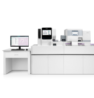

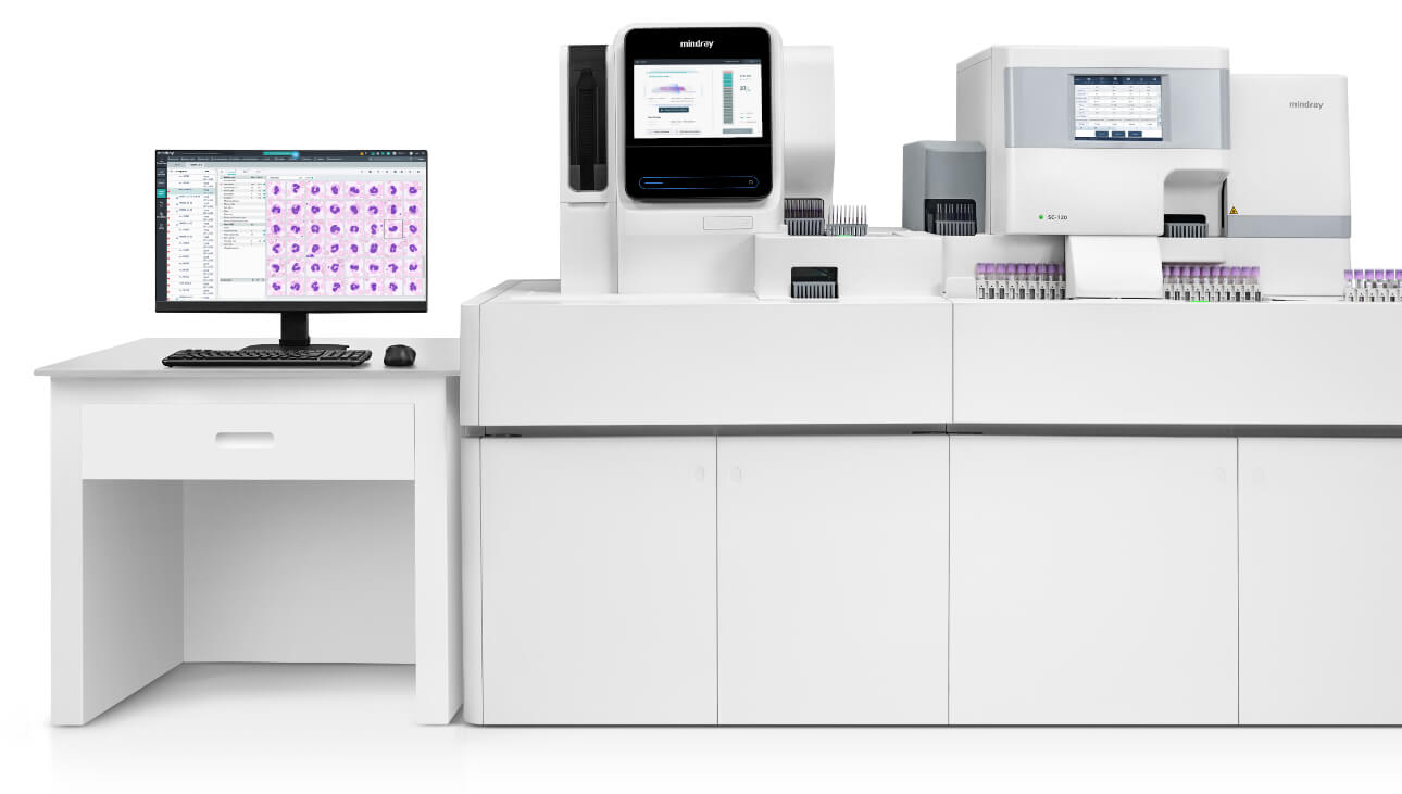
Facilitation of cellular analysis through technological advancements in hematology
Mindray 2022-12-27

Dr. Anil Handoo, Sr. Consultant & Director - Laboratory Medicine (Pathology) Nanavati Max Super Speciality Hospital, Mumbai, is an expert for various national and international committees for hematology and flow cytometry, and also faculty – Flow Cytometry for industry and academic institutions. His areas of expertise are hematopathology, Flow Cytometry, Hematologic Disorders, Coagulation, and Cytomorphometry. He is also a technical assessor for NABL in the fields of Hematology, Molecular Hematology and Flow Cytometry.
He has more than a decade of experience in the overall operation and administration of a laboratory with competency in standardization and quality control of laboratory procedures and expertise in reporting and providing consultation for malignant and non-malignant hematological disorders. In addition to a diagnosis of hematological malignancies by flowcytometric immunophenotyping, stem cells, molecular hematology, and “thalassaemia-screening” utilising HPLC/hemoglobin electrophoresis, working on and optimization of hematology analyzers are close to his heart and form his core interest with a number of publications to his credit.
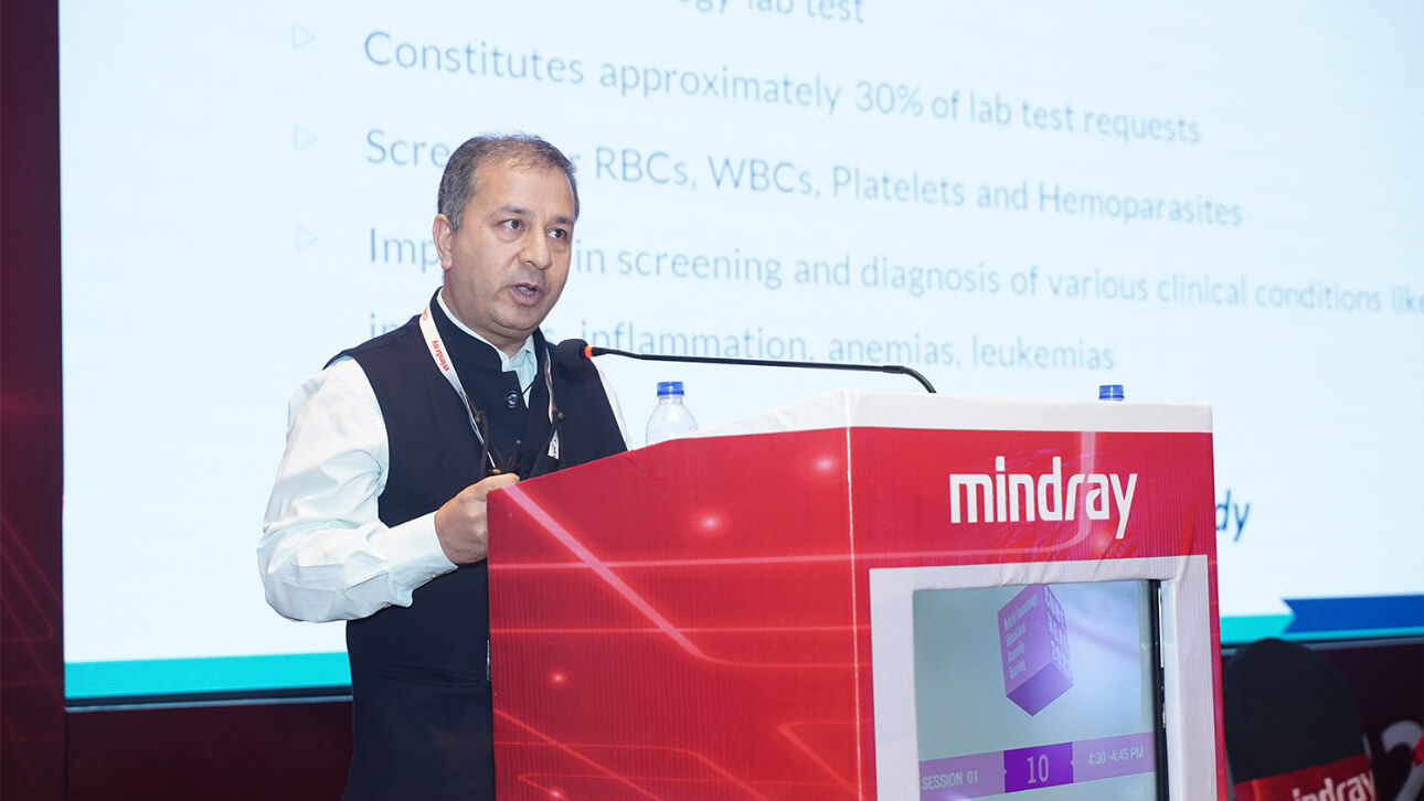
When asked about the aspects of digital Morphology that need to change or evolve, he opines that change is constant, and we habitually settle for that.
Dr Handoo strongly believes that High-speed scanning with the upgradation of AI and continuous machine learning for the pre-classification of cells would help improve the output. “Also, remote web-based access with cloud-based data storage is the way to help us utilise the system better,” says the expert.
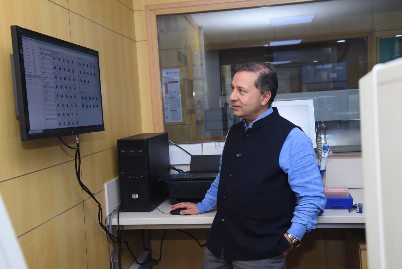
Dr. Anil answered a few questions at the conference regarding the changes and technology being adopted in the field of his expertise and how technologies ensure effective results.
Q. You have almost two decades of experience in the field of Hematology. According to you, what are the major factors that have changed the face of Hematology today?
Recent years have witnessed remarkable strides in hematology analyzer technology. Automation of critical steps and technological
advancements have revolutionized cellular analysis, optimizing lab workflows. Modern analyzers, enhanced by improved flag optimization, not only differentiate mature cells but also detect abnormal cells accurately. The incorporation of Digital Morphology and Artificial Intelligence (AI) with Machine Learning algorithms has taken this further, enabling precise detection of incidental findings that could be overlooked in manual microscopy due to time constraints. Hematology analyzers redefine efficiency and accuracy in lab work.
Q. If you have to explain the concept of Digital Morphology to a layman, how will you do that?
Digital Morphology is nothing, but visualization of peripheral blood smear microscopy using a camera, and automating the image capture with projection onto a computer, instead of manually seeing a slide on the microscope. Digitization and AI-based pre-classification of cells on the smear ensure less amount of time spent on every smear with enhanced workflow efficiency. There is also an advantage of having stored digital image data not only for reviews at a later date but also a possibility of remote viewing and opinion seeking.
Q. How has Digital Morphology revolutionized the field of Hematology?
Digital Morphology, with AI-based algorithms, has ensured highly reproducible cellular classification, and quantification of abnormalities, not only for white blood cells but also for red cells and platelets. It has changed the realms of cellular analysis. The possibility of scanning the smear on the edges for the presence of platelet clumps, or any other abnormalities enhances pick-up rates of such abnormalities. Additionally, the review and valued opinion of an expert from a remote location, with real-time incorporation into the report has been made possible.
Q. What is the role of AI in Digital Morphology? Can you explain with one/two examples?
In the world of hematology, AI is the game-changer, expanding the horizons of morphology analysis. Hematology analyzers with predictive AI capabilities are scaling new heights. Picture this: even the tiniest abnormal cells or parasites, often missed by the human eye, stand little chance of escaping the scrutiny of a digital morphology analyzer. Hematology analyzers emerge as the unsung heroes, spotlighting the minutest anomalies for precise detection and analysis.
Q. Will AI replace the role of humans in pathology laboratories?
I do not see that happening in the near future. However, AI-assisted cell analysis helps humans in obtaining more precision and accuracy.
Q. To what extent can we rely on the results delivered by AI?
As long as the instrument is well-maintained and the smear is appropriately stained, I would trust AI to do a reasonably good job most of the time.
Q. How can Digital Morphology be made affordable so that it can reach the masses? What are your suggestions?
Increasing the awareness regarding use of Digital Morphology would ensure increased demand. In parallel, the miniaturization of devices with smaller footprints and reduction of production costs would help make them affordable to the masses.
Q. According to you, what are the aspects of Digital Morphology that need to change or evolve?
High-speed scanning with the upgradation of AI and constant machine learning for the pre-classification of cells would help improve the output. Also, remote web-based access with cloud-based data storage is the way to go to help us utilize the system better.
Q. Can you please share your experience of using Mindray’s Haematology System in your laboratory?
The adoption of newer methodologies gets you across newer challenges; Digital Morphology Platform (DMP) integration into our routine workflow was no exception. Our initial experience has been that of learning and change management. However, once we settled, the experience has been exhilarating. We have had some very interesting and beautiful pick-ups by the DMP, and many beautiful images of
cells with characteristic morphologies. While we bask in the glory of digital morphology, I still feel there are some areas of improvement and I trust Mindray Team is constantly working to improve the system.