This blog explores the key characteristics and clinical applications of various transducer types. Topics include:
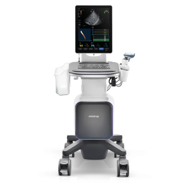
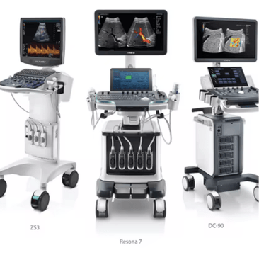
Selecting the appropriate ultrasound transducer directly influences diagnostic image quality, with linear transducers offering high-frequency imaging (5-15 MHz) for superficial structures, curvilinear transducers providing deeper penetration (2-5 MHz) for abdominal assessments, and phased array transducers enabling cardiac evaluations. Understanding transducer characteristics supports diagnostic accuracy across clinical applications. Mindray's ultrasound systems feature comprehensive transducer portfolios for point of care and diagnostic settings.
Ultrasound imaging continues to gain popularity and is quickly becoming the imaging modality of choice for a variety of clinicians, ranging from Emergency Medicine, Critical Care, Radiology Labs, Vascular Labs, Ambulatory Care Centers, and more. Ultrasound is a cost-effective, real-time, and patient-friendly imaging alternative that can be done practically anywhere. These attributes have helped fuel ultrasound’s popularity across many medical fields and specialties. With ultrasound gaining popularity, it has raised questions about the many transducer types and offerings accompanying ultrasound machines.
What Do Ultrasound Transducers Do?
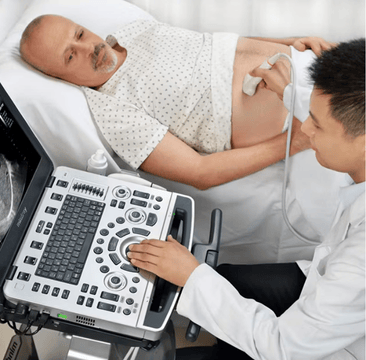
Ultrasound transducers are used to image various body parts. To use an ultrasound machine, you need to have a transducer that connects to the ultrasound machine. Once the transducer is connected to the ultrasound machine, the transducer is placed on the patient, and the clinician can visualize images of the patient’s anatomy.
Transducers connect to the ultrasound via ports. Transducer ports are traditionally found at the bottom of the ultrasound machine if the system is cart-based. For laptop-based systems, the transducer ports are located on the right and left sides of the laptop. For tablet-based ultrasound machines, the transducer ports are typically found behind the tablet user interface.
What Is an Ultrasound Transducer Made Of?
Transducers are made of a plastic or metal housing, protecting the internal electrical components.
How Do Ultrasound Transducers Work?
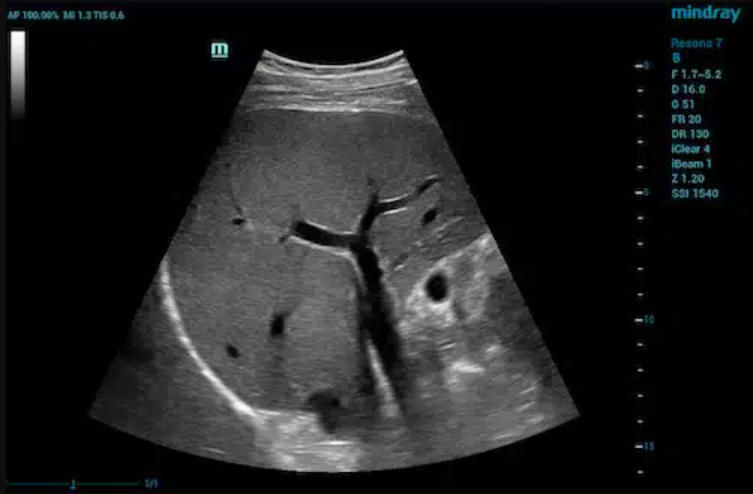
Ultrasound transducers transform electrical energy into mechanical energy. The electrical signal from the ultrasound machine is transformed via piezoelectrical crystals inside the transducer into a mechanical sound wave. These waves permeate the body, reflect off of internal structures, and return to the transducer. There the process is reversed, and the transducer changes the returning sound waves into electrical signals, which are then interpreted and displayed as images on the ultrasound screen. Additionally, materials are added to the housing (known as backing) and matching layers.
What Types of Transducers Are Used in Ultrasound?
There are three main types of transducers – convex/curvilinear, linear, and phased/sector transducers. In addition, there are many specialty transducers, such as endocavity, transesophageal, volume, and continuous wave (CW)/pencil transducers. Different types of transducers are used for different ultrasound exams and are ideally suited to image different structures.
Main Types of Transducers
Convex/Curvilinear Ultrasound Transducers
A convex transducer is a curved transducer. The shape of a convex transducer determines the sector width and shape of the image. A convex transducer offers a wider field of view for larger or deeper structures. Convex transducers are most frequently used for abdomen, obstetrics/gynecology (OB/GYN), urology, and some musculoskeletal (MSK) applications.
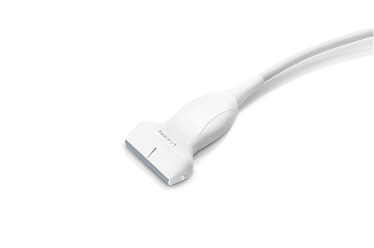
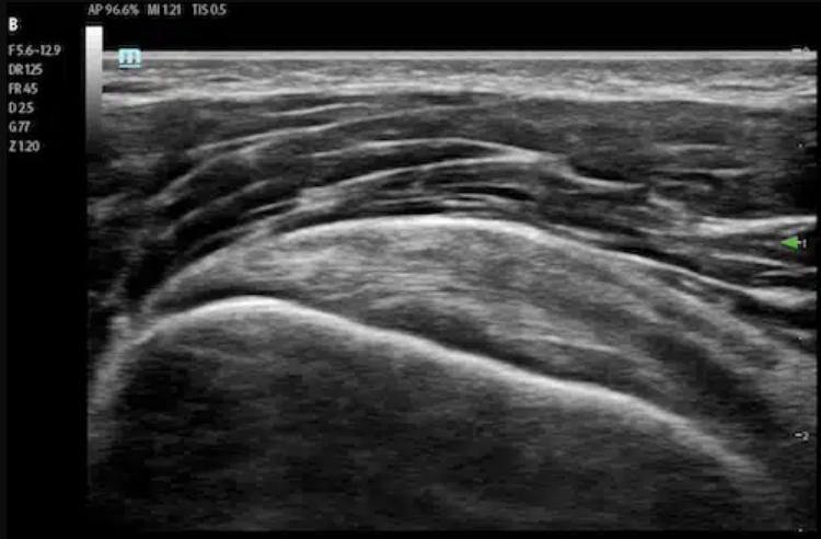
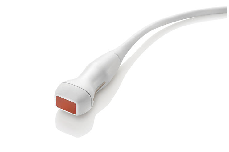
Linear/Sector Ultrasound Transducers
A linear transducer is a straight transducer. The linear transducer’s length determines the image’s sector width and shape. A linear transducer offers detailed resolution at superficial depths. Linear transducers are most frequently used with MSK, nerve, small parts, vascular, and pediatric applications.
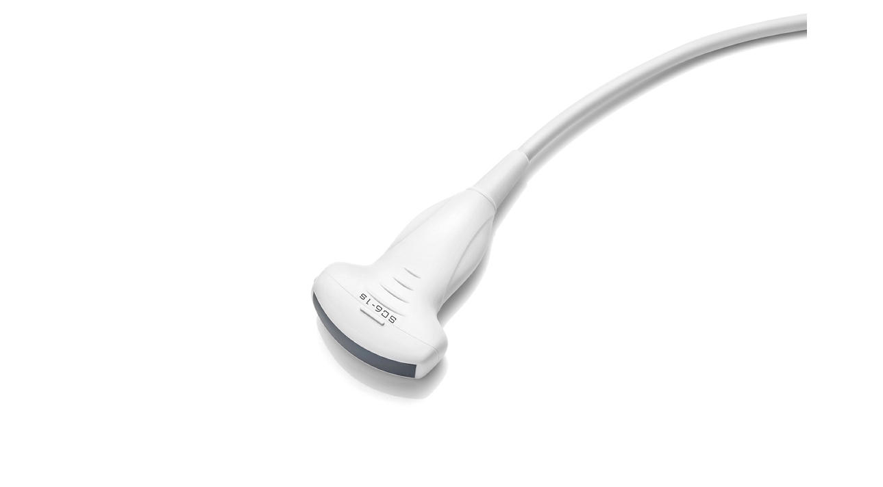
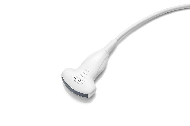
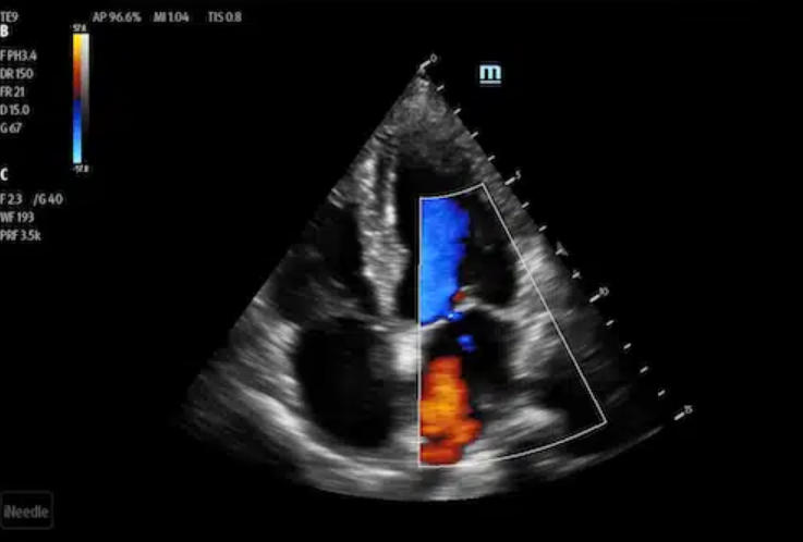
Phased Ultrasound Transducers
A phased transducer has a small footprint with a sector image shape and features high temporal resolution and penetration. This allows clinicians to image structures that are moving in real-time. Phased transducers are most frequently used with applications ranging from cardiac, transcranial, abdomen, and pediatrics.
Specialty Transducers
Endocavity Transducers
An endocavity transducer is a specialty transducer used to image structures from inside the body. This allows for better visualization of structures that are not easy to view with a surface transducer. The shape of the imaging surface provides a very wide field of view. Endocavity transducers are most frequently used for OB/GYN and urology applications.

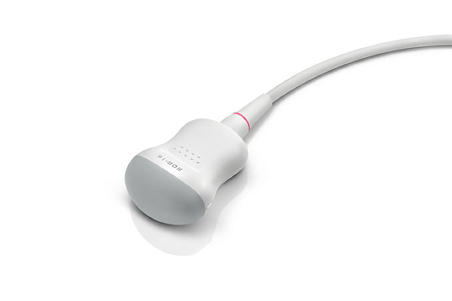
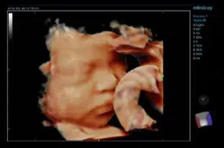
Volume Ultrasound Transducer
A volume transducer is a specialty transducer that captures 3D/4D images and can be utilized in a variety of applications. They can be useful in helping clinicians to appreciate depth better and achieve a more realistic representation of anatomy in additional planes. Volume transducers are typically used for cardiac and OB/GYN imaging.
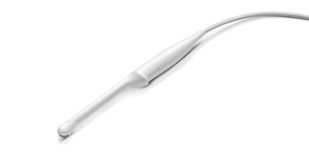
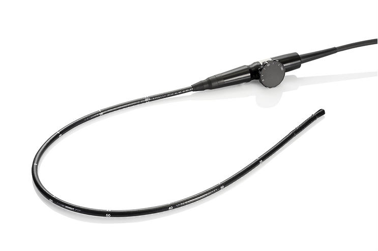
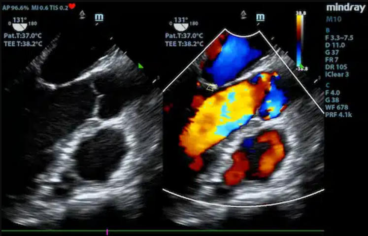
Transesophageal Echo Transducers
A transesophageal Echo (TEE) transducer is a specialty transducer used to image structures from inside the body. It is designed to be inserted into the esophagus and image the heart from behind. This offers the ability to image structures that cannot be well visualized in a transthoracic echo – TEE imaging offers an unobstructed view of the heart (i.e., lungs and ribs do not get in the way). The image will be a sector shape that offers a wide field of view. TEE transducers are used exclusively for cardiac imaging.
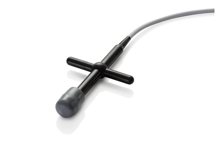
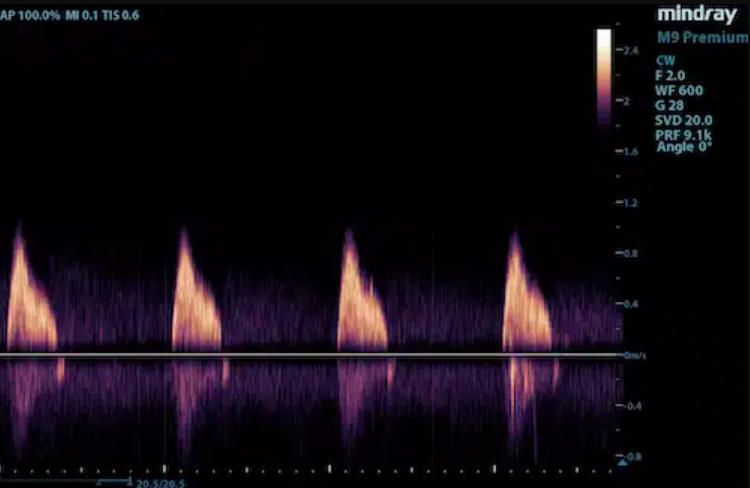
Continuous Wave (CW)/Pencil Ultrasound Transducers
A Continuous Wave (CW) transducer has a circular-shaped tip. These non-imaging transducers are dedicated to Doppler interrogation to help clinicians see waveforms and hear blood flow. CW transducers are used for vascular, cardiac, and transcranial applications.
The Mindray Difference
Clinicians rely on tools and technologies to assess patients quickly and reliably. At Mindray, we believe all clinicians deserve cost-effective, reliable medical devices that empower them to provide the best possible care for their patients. To ensure we continue to develop the tools clinicians need today and, in the future, we accompany and listen to customers and create a collaborative experience that results in innovative solutions that address the challenges they face every day. Experience peace of mind and see something better with Mindray Ultrasound.
Browse Our Ultrasound Solutions