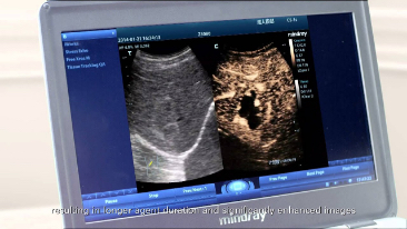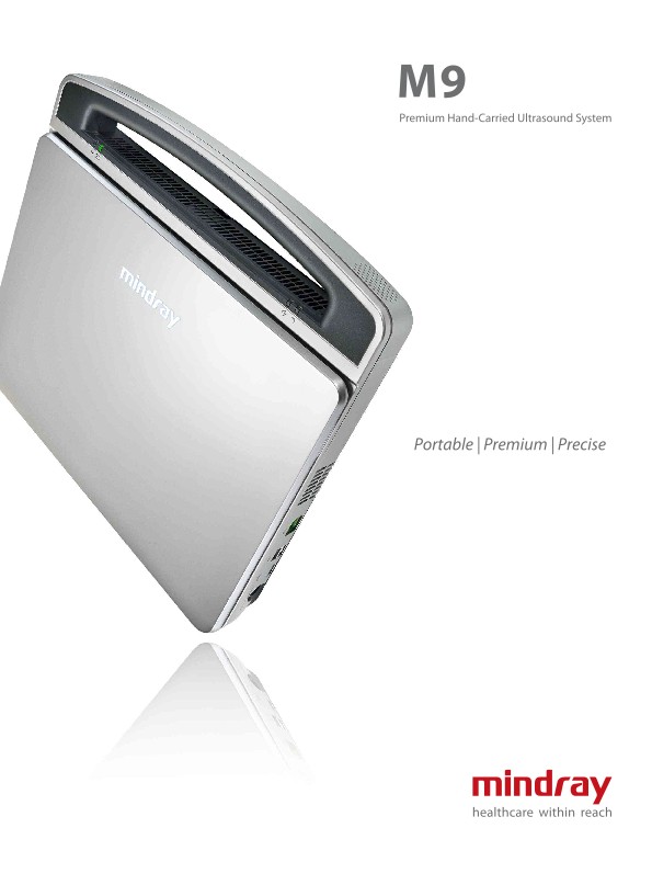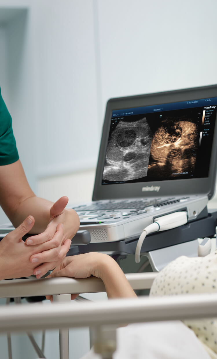
Based on Mindray's new generation ultrasound platform, mQuadro, M9 has raised industry standards to a whole new level. Advanced signal transmission and reception processors provide highly sensitive and accurate echo detection. Innovative transducer technologies allow for better penetration and higher resolution to enhance your diagnostics.

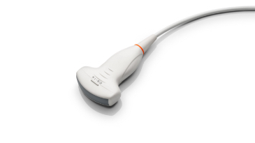
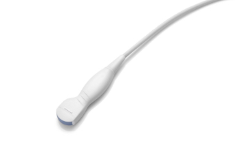
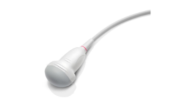
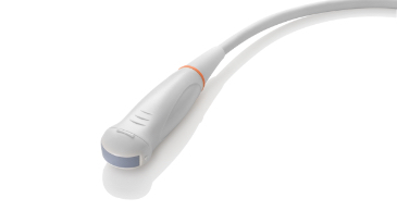
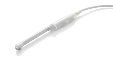
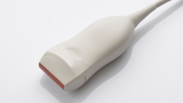
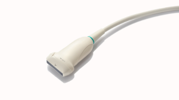
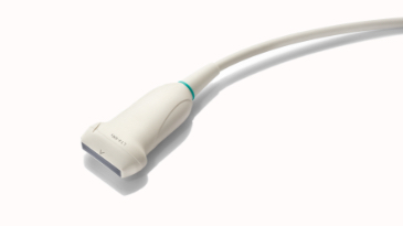
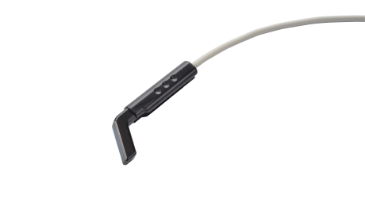
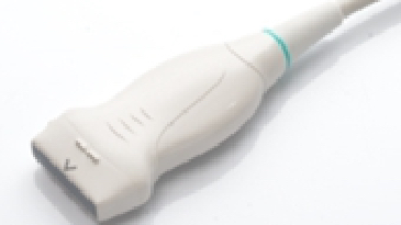
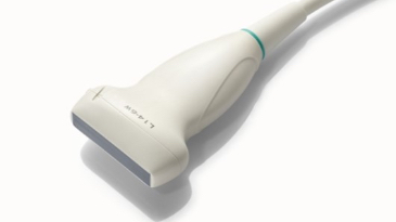
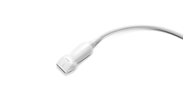
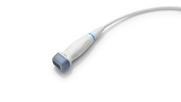
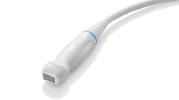
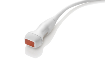
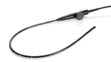
3T Transducer and Single Crystal Technologies
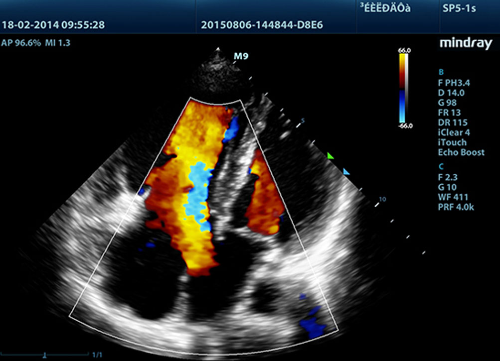
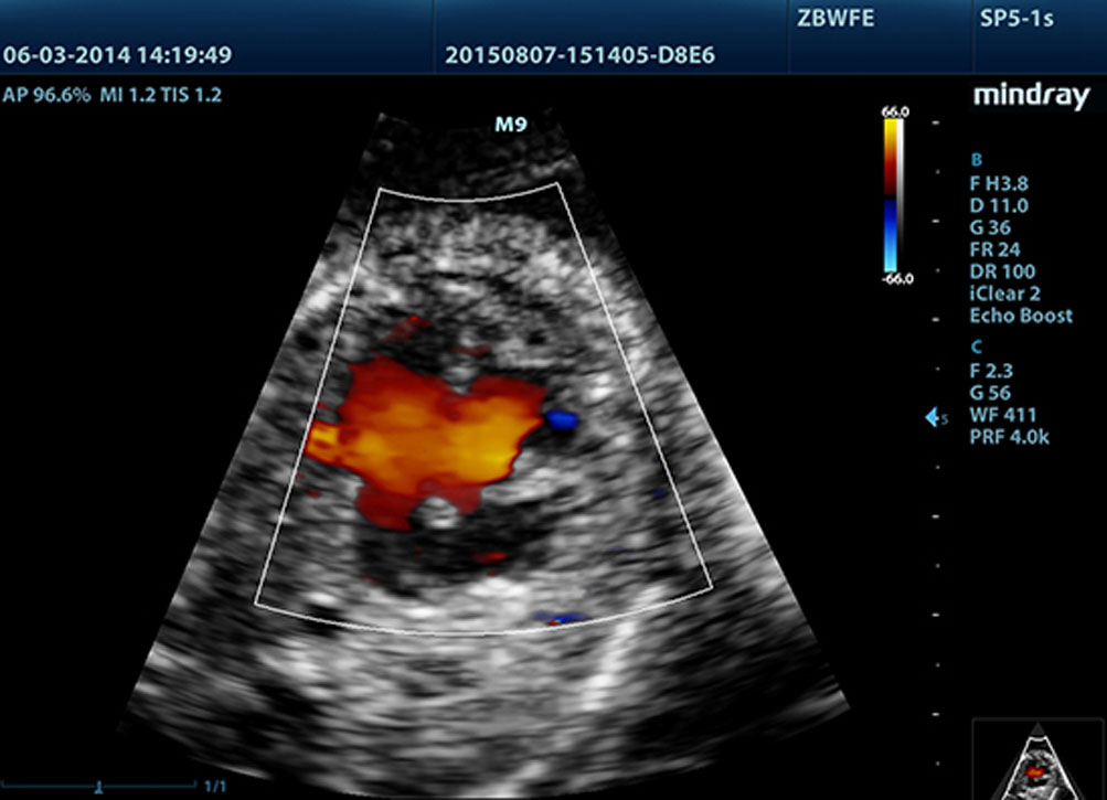
Providing sharper images, all probes compatible with the M9 come equipped with Mindray’s unique 3T transducer technology. Enhanced with the addition of single crystal technology, M9 offers better penetration and color dynamic flow, especially during difficult-patient scanning.

Echo Boost™
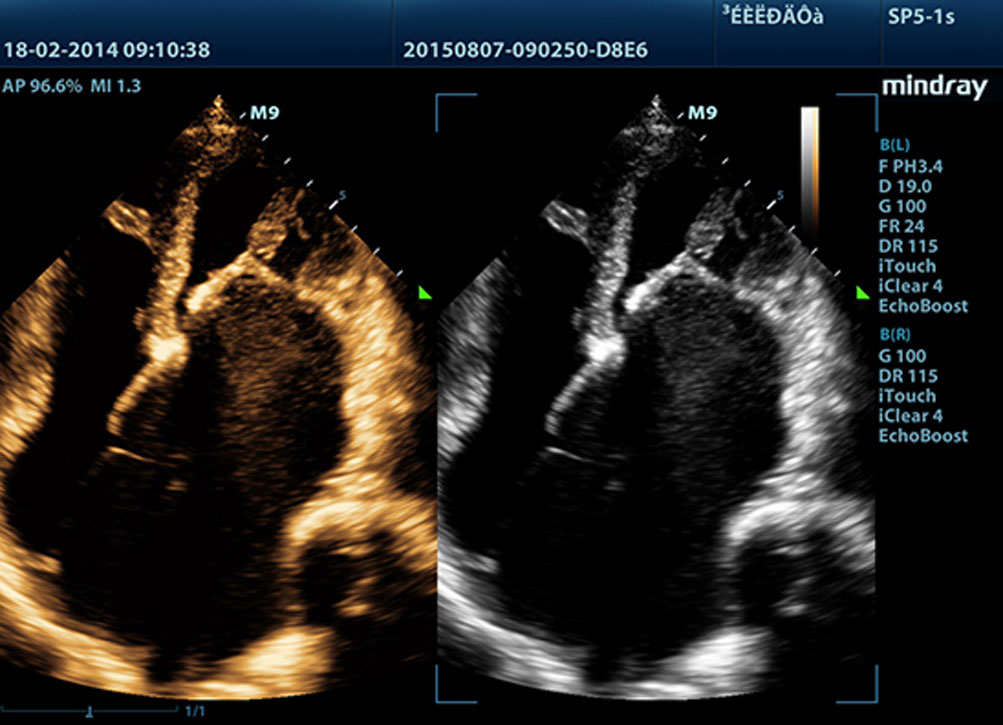
Mindray’s unique adaptive signal processing technology and intelligent echo detection is designed to use native signal-to-noise information to enhance weak echo signals while suppressing clutter noise. This results in more balanced image brightness and improved visualisation of myocardium tissue layers.
Tissue Tracking with Quantitative Analysis
The TT QA functionality on M9 allows for a simple, quick and non-invasive evaluation of left ventricular wall motion abnormalities. Supported by Mindray’s patented 3T technology with single crystal, M9 controls the image drift caused by probe movement and breathing to significantly improve tracking accuracy and effectiveness. With the added unique benefit of on-site analyses, the TT QA on M9 can be performed at the bedside, saving time and making complicated diagnoses much simpler.
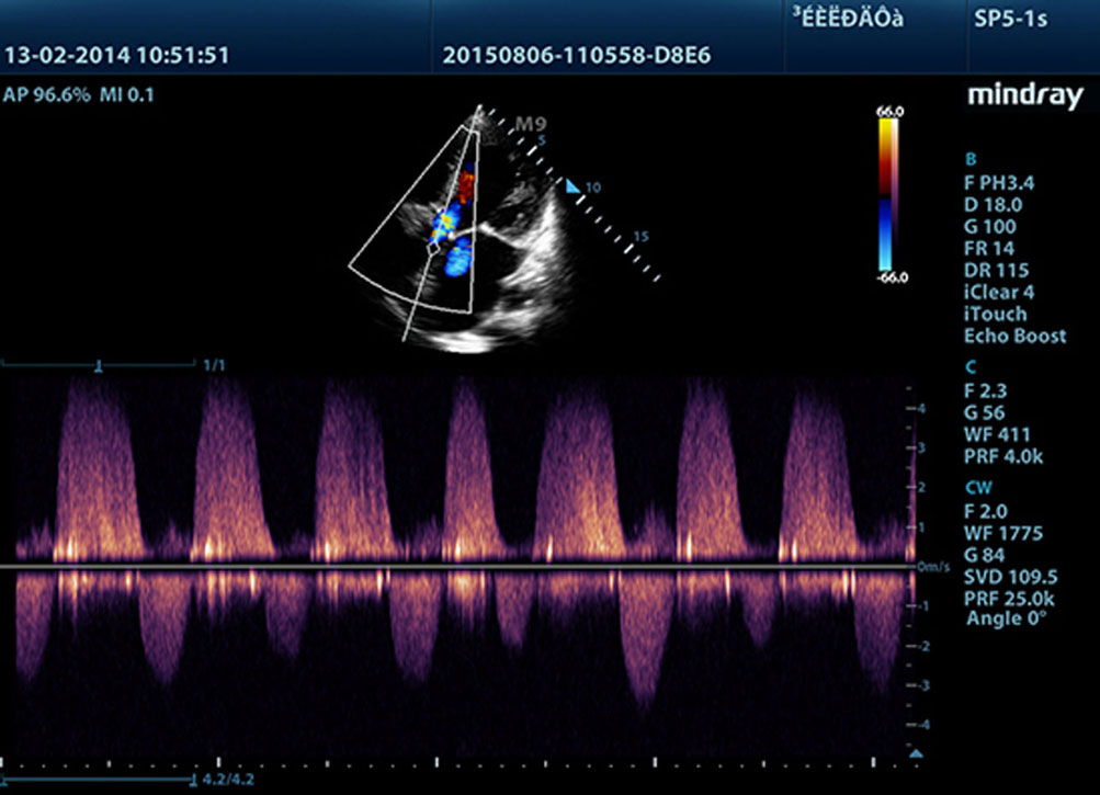
LVO with Stress Echocardiography
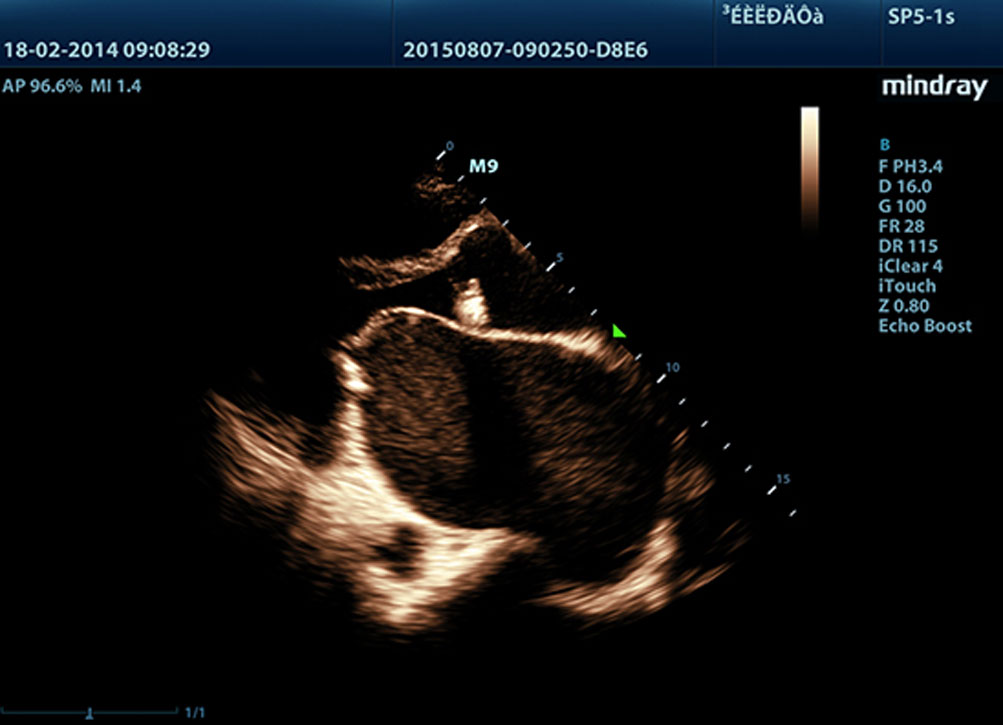
M9’s premium capabilities allow for LV opacification during stress. With enhanced discrimination between myocardial tissue and blood pool, practitioners can get better visualisation of the endocardial surface. The Stress Echo feature on M9 includes a complete package for pharmacological stress and exercise stress echo. The package is supported by a flexible reporting system that can be optimised for your individual needs.
PSHI™ (Phase Shift Harmonic Imaging)
Purified Harmonic Imaging offers better contrast resolution and clearer images with less noise.
Tissue Harmonic Imaging (THI)
Using second harmonics generated from tissue boundary layers, THI significantly enhances contrast resolution and improves image quality for technically difficult subjects.
Tissue Specific Imaging (TSI)
Tissue Specific Imaging optimises the image quality based on the properties of the tissue being scanned. Four imaging options are available including general, muscle, fluid and fat.
iBeam™
Scan from multiple angles to form a single image for enhanced contrast resolution and improved visualisation.
iClear™
Gain improved image quality based on auto structure detection:
- Sharper and continuous edges
- Smooth uniform tissues
- Cleaner ‘no echo areas’
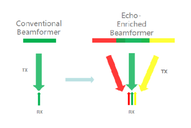
Echo-Enriched Beam Forming
Traditionally neglected echo signals of adjacent beams are formed into one finer, stronger imaging beam. This provides better ‘out-of-focus’ image resolution and deeper image penetration.
Multi-Beam Formation
Up to 12 beams can be transmitted at once, resulting in excellent time resolution and higher frame rate.

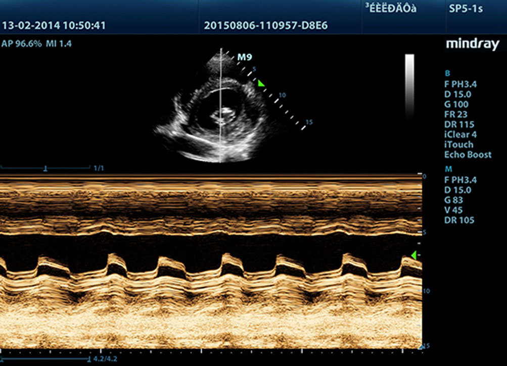
Free Xros M™
Gain precise anatomical observation by freely placing sample lines at any angle. Obtain better images through simultaneous display of up to 3 sample lines.
Free Xros CM™
Accurately evaluate myocardial motion at different phases and simultaneously determine myocardial synchronisation. The high frame-rate provides you with accurate results.
Auto EF
Automatic recognition of the diastole and systole from 2D echo clips and output EDV/ESV/EF etc. results using the Simpson method.
TDI
Tissue Doppler Imaging allows you to quantitatively evaluate local myocardial movement and function, providing complete TDI modes for faster and accurate diagnoses.
Workflow
iZoom™
Set a full screen view with a swipe.
Auto IMT (Intima-Media Thickness)
Auto measurement of anterior and posterior wall thickness provides an accurate carotid status.
iStation™
Mindray’s unique Patient Information Management System lets you integrate, review, archive and retrieve patient data effectively.
iTouch™
Gain instant auto image optimisation in B, Color and PW Modes with the touch of a single key.
iWorks™
A smart tool to let you focus more on the patient. This can significantly reduce the patient scan time through standardisation and user-defined capability.
Raw Data
Optimum flexibility for post-processing of stored images including parameter adjustments, adding comments and measurements, to maximise productivity during scanning.
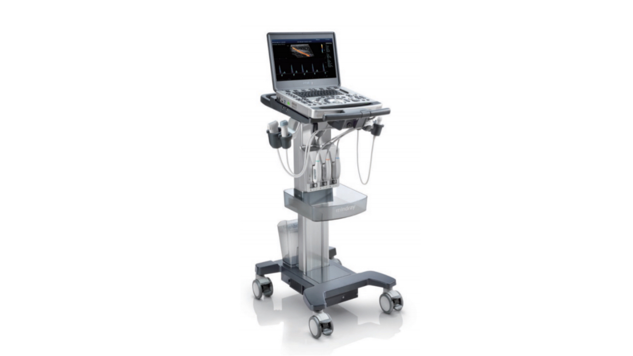
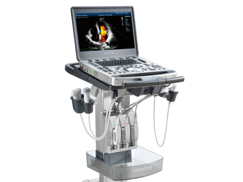
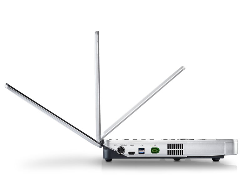
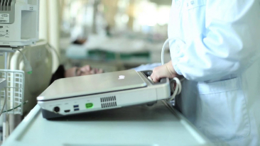
Innovative Unit
· Thin magnesium-alloy body
· 15.6” LED HD monitor with slim design
· Built-in battery providing 90 minutes scanning time
· High capacity SSD hard drive for more secure patient data
Customised Trolley
· Inbuilt quick and easy locking system
· iPower: over 3.5 hours scanning with trolley mounted battery pack
Green All The Way
· Noiseless system
· Automatic brightness adjustment
· Reliable RoHS certified materials