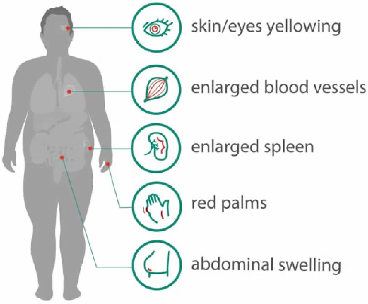
If you are suffering from abdominal swelling and enlarged spleen, or find unusual physical performance such as enlarged blood vessels beneath the skin, red palms, yellowing of your skin and eyes, be cautious, you might have Non-Alcoholic Fatty Liver Disease (NAFLD).
A Growing but Silent Killer
As the name implies, NAFLD is an umbrella term for a range of liver conditions affecting people who drink little to no alcohol. Its main characteristic is too much fat stored in liver cells.
What’s worse? Without timely monitoring and proper interfering, NAFLD is likely to progress into nonalcoholic steatohepatitis (NASH), which is a more severe condition that involves inflammation of the liver and possibly cell damage which can lead to liver fibrosis, or scarring of the liver, resulting in higher possibility of dying.
Therefore, it is of great importance to detect and assess the stage of liver fibrosis in people suffering from NAFLD early to prevent further progression or serious consequences.
Accurate Evaluation Brings Better Aid
Take a 45 years old man’s case as an example. Suffered from the abdominal pain, the man looked for help from a team of experts in Italy.
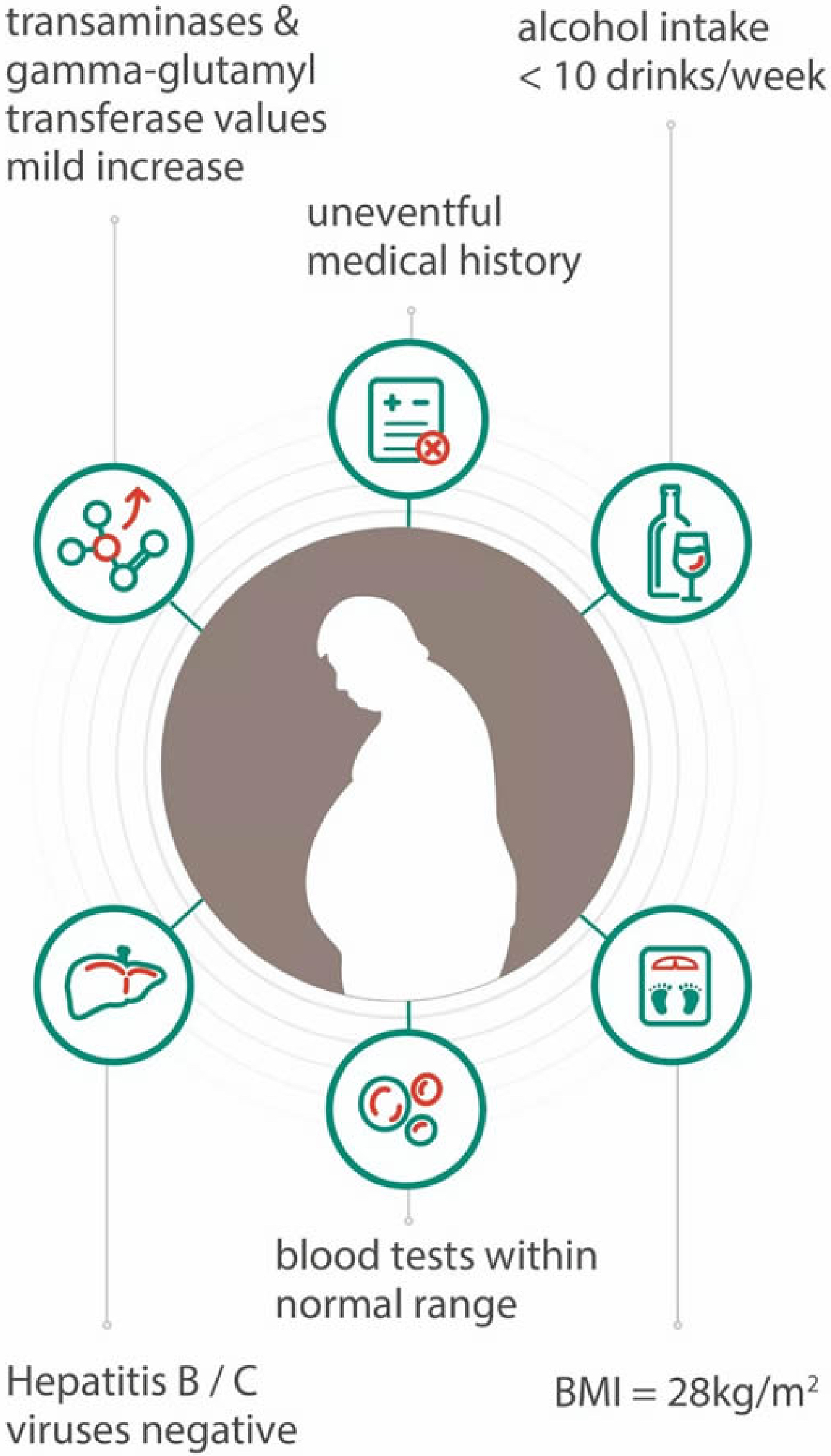
After simple examination, experts found out that he was overweight with a BMI of 28kg/m^2, a weekly alcohol intake of less than ten drinks and an uneventful past medical history except for an appendectomy at the age of 12. His platelet count and other blood tests were within the normal range and he came up negative for Hepatits B or Hepatitis C viruses. However, there was a mild increase in his transaminases and gamma-glutamyl transferase values.[1]
To better understand his condition, the team progressed into a non-invasive, rapid and accurate method, the ultrasound examination.
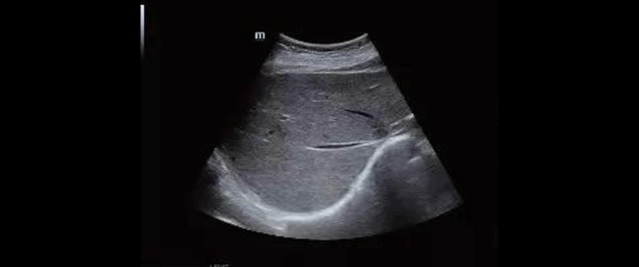
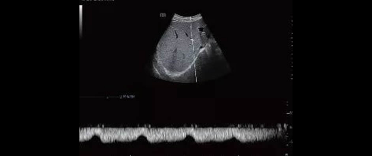
To their surprise, a bright liver with smooth margins and patent portal vein has been found using the B-mode and Doppler of Mindray’s Resona 7. Moreover, the mean portal blood flow velocity was 28 cm/s and the flow in the hepatic veins had a biphasic pattern.[2]
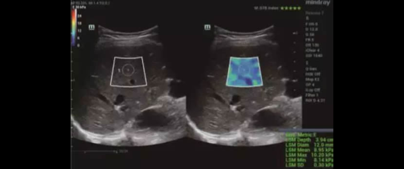
Applying with the Sound Touch Elastography, the system showed a median value of 8.83 kPa (IQR/M = 1.8%) which indicated significant fibrosis. The liver stiffness measurement was also reliable.
Transient elastography with a FibroScan device was performed in the same session soon after the evaluation with the Resona 7 and the values obtained was 8.5 kPa. Liver biopsy, performed a week later, showed stage 2 fibrosis.[3]
With availability of an ultrasound system, in which shear wave elastography technique is implemented, experts were able to evaluate the liver stiffness whose results were compatible with significant fibrosis (F2), confirmed by liver biopsy later.
With a clearer picture of his condition now, the team can deliver better aid for him.
Navigate Stiff Tissue is the Key
As the case implied, STE can significantly increase the odds of early detection by creating a safe ultrasonic shear wave to disturb soft tissue as it penetrates through the tissue. The ultrasound system can then analyze and evaluate tissue stiffness within a selected region in the scanning area.
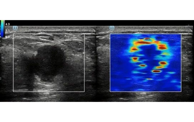
After that, it will interpret and display real-time information in a color-coded image based on the softness and stiffness of the tissue which help to indicate what kind of anomaly is present.
For example, with STE, the possible presence of liver fibrosis can be detected for further follow-up or rule the possibility out by not seeing any stiff tissue.
In addition, STE provides a motion stability index and reliability chart to help ensure accuracy and reproducibility of the examination.
“
“In ultrasonography today, we have to combine a perfect B-mode image, a perfect color Doppler image and a perfect STE image in order to have a global overview on what’s happening in the nodule and how suspect benign or malignant this nodule is.” said Pavlos Zoumpoulis, President and CEO of Diagnostic Echotomography SA, Greece
”
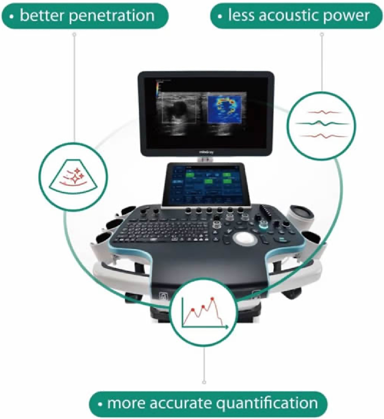
In a nutshell, integrated with Mindray’s exclusive Ultra-wide Beam Tracking technology, STE demonstrates a giant leap on ultrasound performance, mainly manifesting in better penetration, more accurate quantification and less acoustic power.
As the incidence of NAFLD continues to soar, quick detection and evaluation will contribute greatly to improving clinician’s confidence on disease diagnosis and early intervention, benefiting patients in the end.
With profound insights into such crucial clinical needs, Mindray will continue to push medical boundaries, bringing better healthcare within reach for everyone.
References:
[1] Ferraioli, Giovanna, MD, Laura Maiocchi, MD, Carlo Filice, MD, and Kendy C. C. Ng, BSc, RDMS, RDCS, RVT, MBA. “Shear Wave Elastography for the Assessment of Diffuse Liver Disease: Protocol and Case Studies.” Ultrasound Unit of the “Fondazione IRCCS Policlinico San Matteo”, Medical School University of Pavia, Italy,2018
[2][3] “Shear Wave Elastography for the Assessment of Diffuse Liver Disease: Protocol and Case Studies.” Ultrasound Unit of the “Fondazione IRCCS Policlinico San Matteo”, Medical School University of Pavia, Italy,2018